

What exactly is a mast cell?
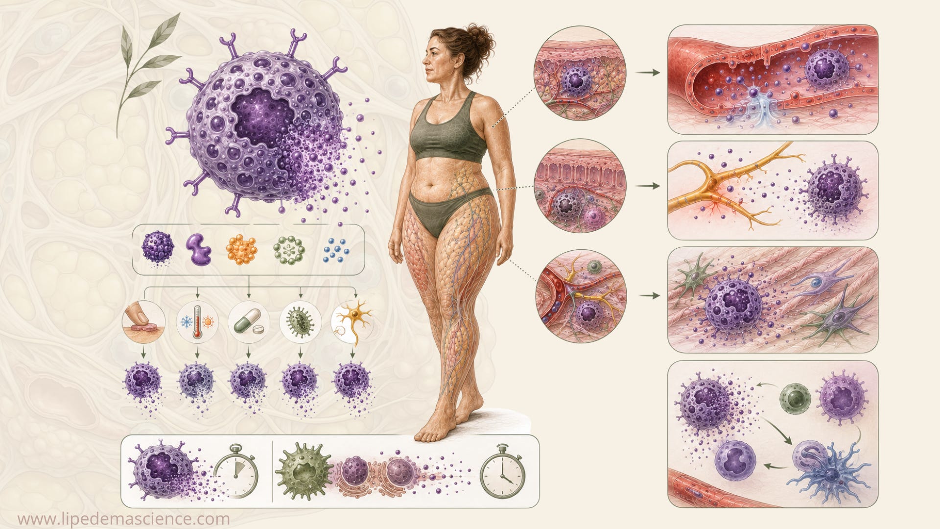
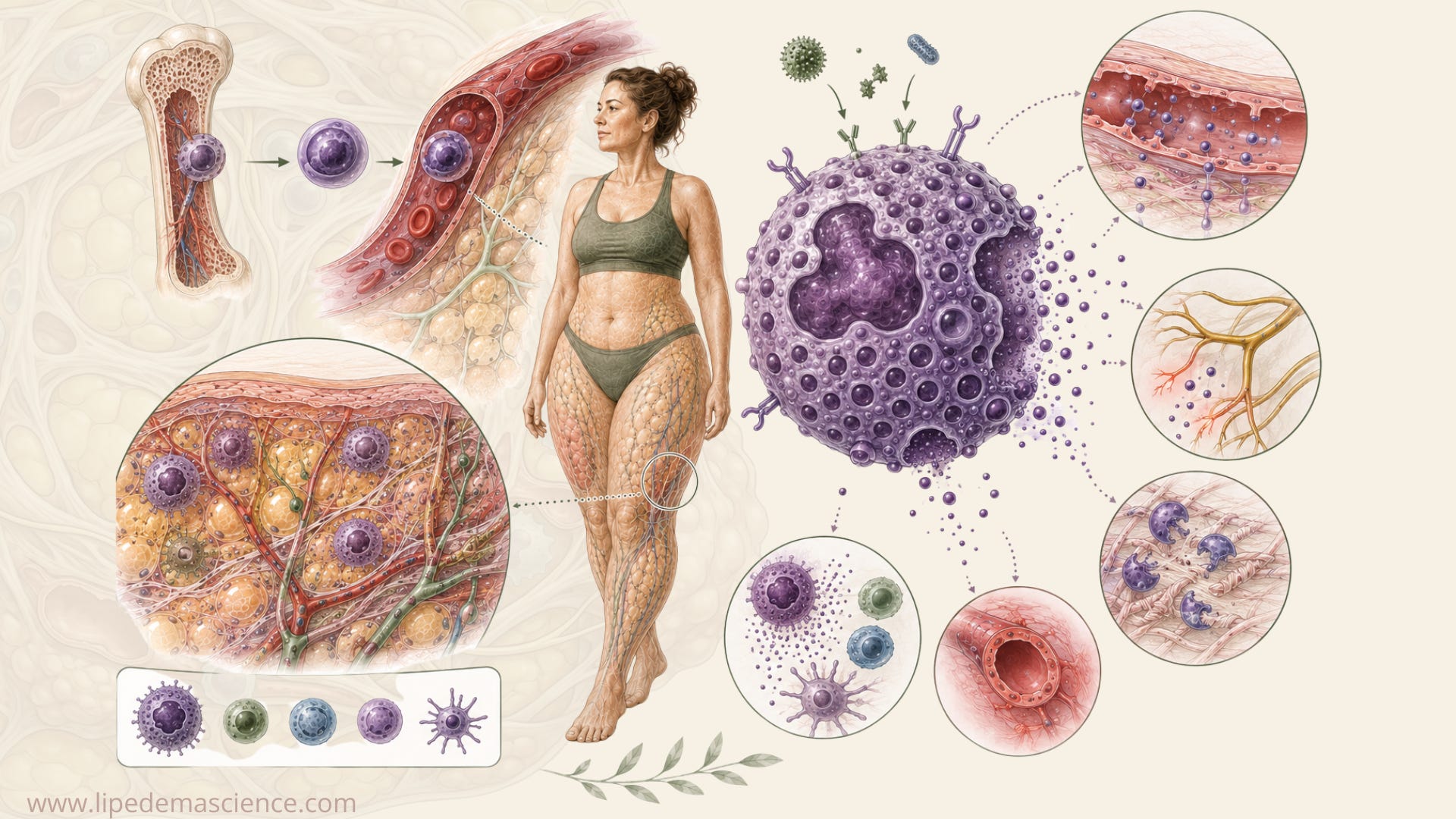
Mast cells are immune cells that live mainly in tissues rather than circulating in the blood. Their precursors originate in the bone marrow and travel through the bloodstream before developing into mature mast cells within tissues. They are especially common in the skin, gut lining and airways, and they are also found close to blood vessels and sensory nerves.
Their location is important. Mast cells are positioned in places where the immune system may need to respond quickly to something happening in the surrounding tissue.
As a LipedemaScience member, you get full access to in-depth articles that translate complex lipedema research into understandable, practical knowledge. I examine new studies, emerging theories and popular claims around topics such as inflammation, hormones, nutrition, pain, lymphatics and treatments, including what the research actually found and where the evidence is still uncertain. Members can also suggest topics they want me to investigate and receive new articles and research updates directly by email.
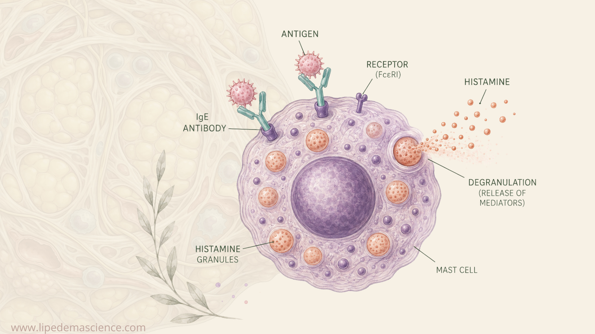
Inside a mast cell are small storage compartments called granules. These contain pre-formed substances, including histamine, tryptase and heparin. When a mast cell receives an appropriate signal, it can release the contents of these granules within seconds in a process called degranulation. This speed is one reason mast cells can produce such immediate effects during an allergic reaction. Some of their most powerful mediators are already stored and ready for release.
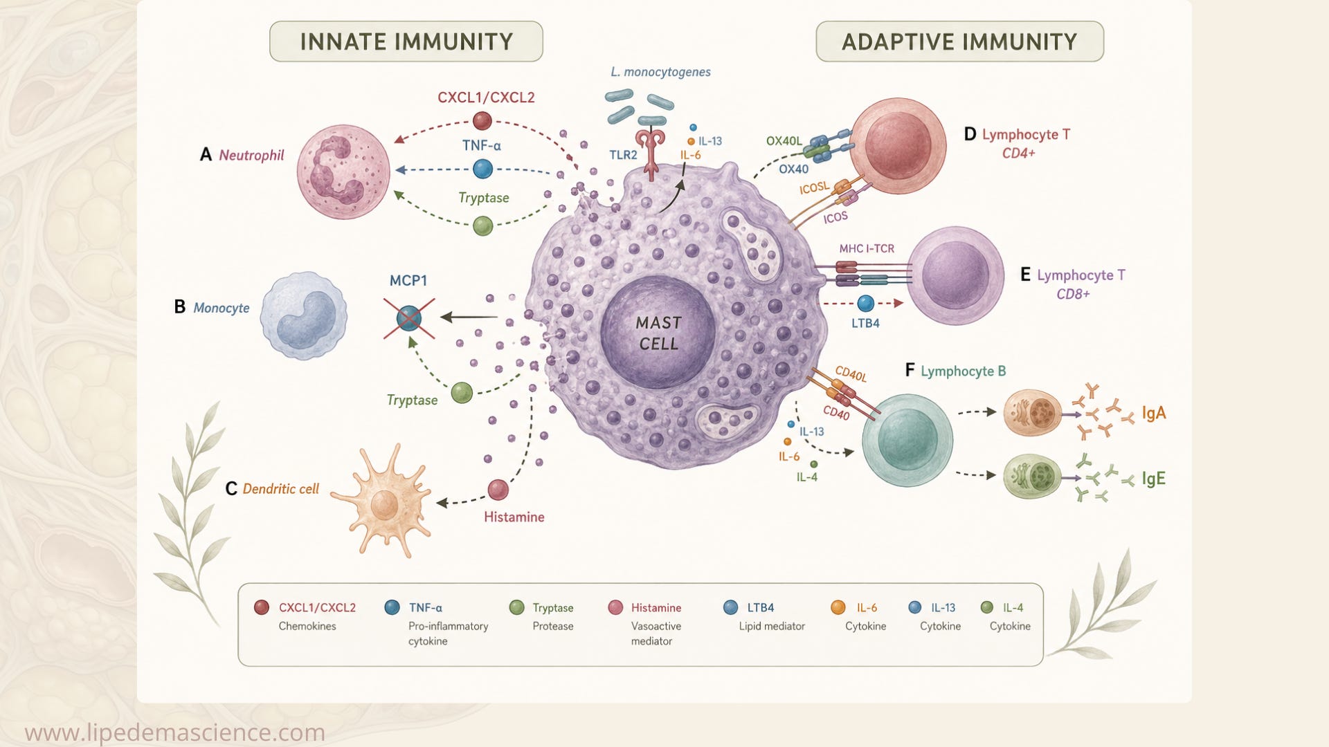
Histamine can increase the permeability of small blood vessels, allowing more fluid and molecules to move into the surrounding tissue. It can also stimulate sensory nerves and contribute to itching and pain. Tryptase and other enzymes can influence the extracellular matrix and tissue remodeling. Mast cells can also produce cytokines, prostaglandins, leukotrienes and other signaling molecules that communicate with immune cells, blood vessels, nerves and connective-tissue cells.
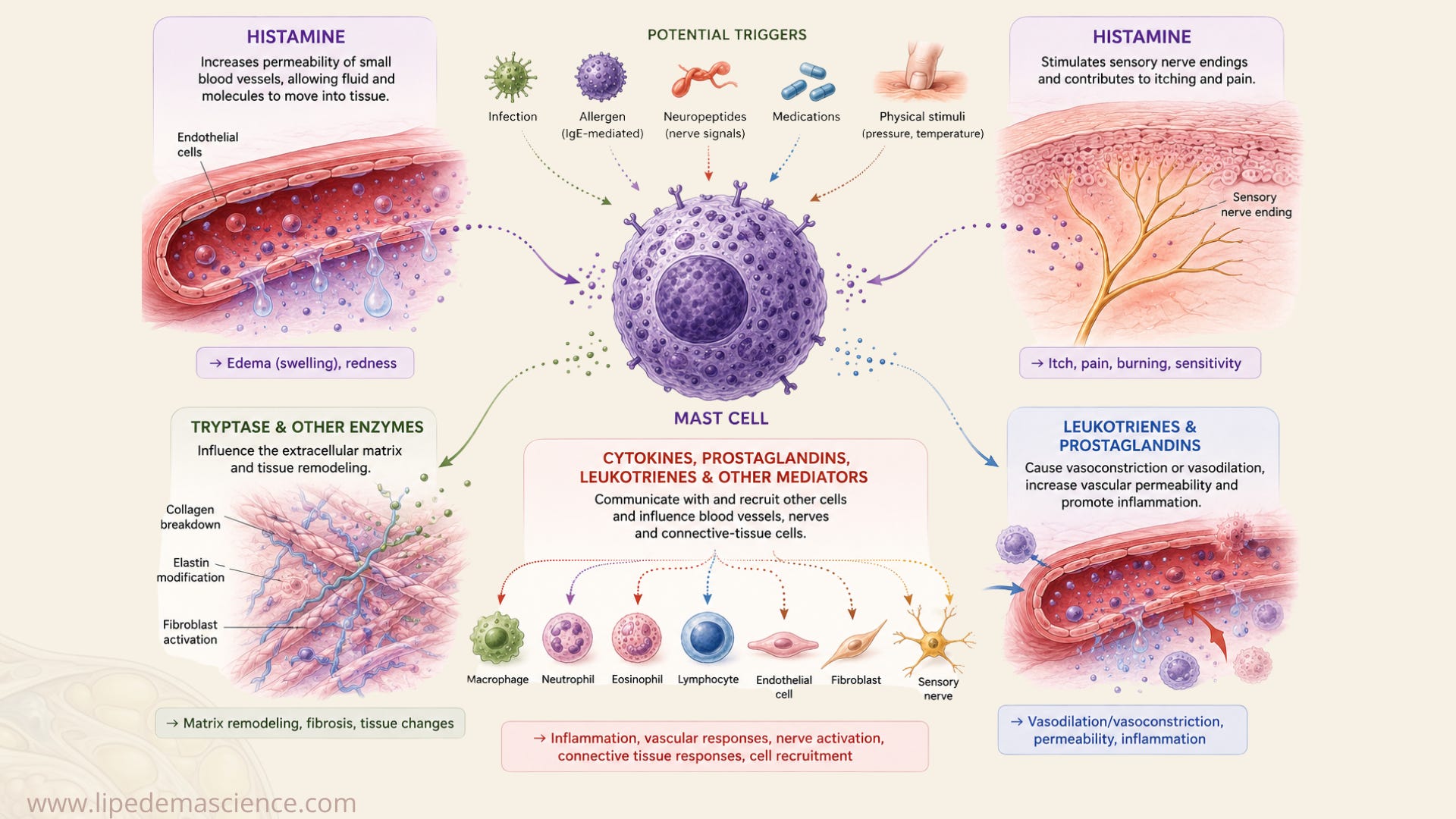
This ability to respond rapidly and influence several biological systems at once is one reason mast cells have attracted so much attention.
Mast cells are not only allergy cells
Allergy is probably the best-known way of activating mast cells. In a classic allergic reaction, IgE antibodies attached to receptors on the mast-cell surface recognize a particular allergen and trigger rapid mediator release.
But IgE-mediated allergy is only one pathway.
Mast cells can respond to signals associated with infection and tissue injury, certain medications, neuropeptides released by nerves and some physical stimuli. Temperature changes and mechanical stimulation can also influence mast-cell responses under particular circumstances.
This versatility is scientifically important, but it also helps explain why mast cells are increasingly discussed as a possible mechanism behind many different conditions.
Mast cells communicate with nerves, blood vessels, connective tissue and other immune cells. Pain, swelling, flushing, itching and gastrointestinal symptoms can therefore all involve biological pathways in which mast cells participate.
This makes mast cells an attractive explanation when several seemingly unrelated symptoms occur together. It also makes them particularly easy to over-attribute.
Showing that mast cells can influence a symptom does not establish that they are responsible for that symptom in a particular disease. Biological plausibility is the beginning of a research question, not evidence that the mechanism has been demonstrated.
Why are mast cells interesting in lipedema?
There are several reasons researchers might want to investigate mast cells in lipedema.
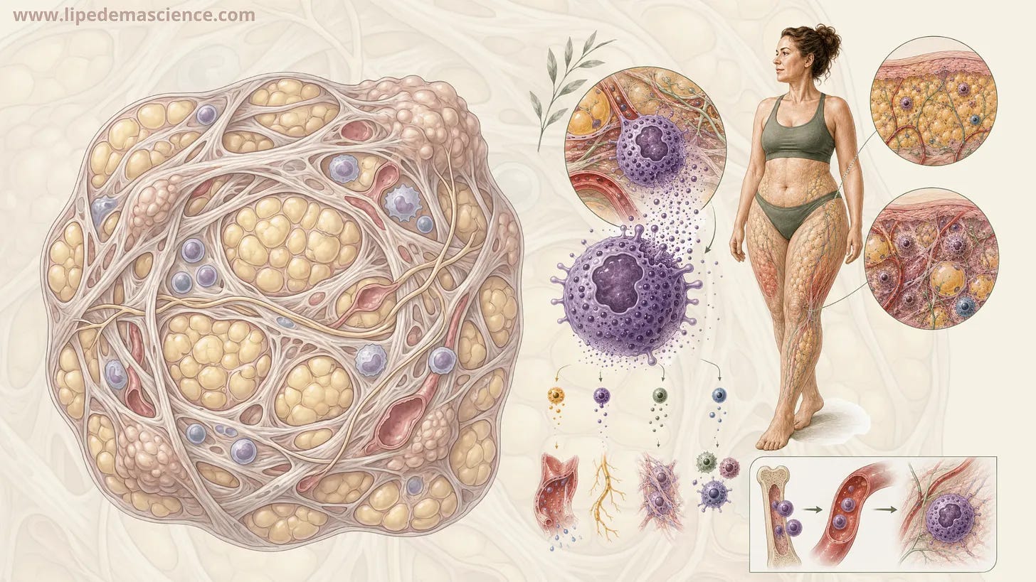
Pain and pressure sensitivity are characteristic features of the disease. Changes involving blood and lymphatic microvessels have been observed in lipedema tissue, and researchers are investigating alterations involving inflammation, connective tissue and the extracellular matrix. Mast cells are capable of interacting with all of these systems.
This creates an appealing biological hypothesis. Mast-cell mediators could potentially influence vascular permeability, sensory nerves, immune signaling and tissue remodeling within lipedema adipose tissue.
The important word here is could.
The question is not whether mast cells are biologically capable of doing these things. We already know that they are. The question is whether abnormal mast-cell activity actually occurs in lipedema and whether it contributes meaningfully to the disease.
That requires research directly involving people with lipedema.
What have researchers actually found in lipedema tissue?
One of the more useful studies for answering this question was published by Al-Ghadban and colleagues in 2019. Researchers examined thigh skin and subcutaneous adipose tissue from women with lipedema and compared it with tissue from controls.
They found several differences in lipedema tissue, including enlarged adipocytes, dilated blood and lymphatic microvessels, increased angiogenesis and increased numbers of macrophages.
The mast-cell result is particularly relevant to the current discussion. The researchers did not establish a significant increase in mast-cell numbers in lipedema tissue compared with controls.
This does not prove that mast cells have no role in lipedema. Counting mast cells and determining what those cells are doing are different questions. A similar number of mast cells could theoretically behave differently or release different amounts of mediators.
But it does mean that claims that lipedema has been shown to involve an accumulation or proliferation of mast cells should be treated cautiously. That has not been established by this comparative tissue study.
What about the study on histamine and mast cells?
Another study, published by Bonetti and colleagues, approached the question differently.
The researchers examined subcutaneous adipose tissue and reported higher levels of histamine and histamine metabolites in samples from women with lipedema compared with controls. They also investigated sodium cromoglycate, also known as cromolyn, a medication with mast-cell-stabilizing properties. After a short treatment period, lower concentrations of histamine were measured in lipedema tissue.
This finding is interesting enough to justify further research, but it needs to be interpreted within the limitations of the experiment.
The treatment component was extremely small and short. It was not a large randomized, placebo-controlled clinical trial, and it did not demonstrate that cromoglycate reduced lipedema pain, swelling, limb volume or disease progression. The primary observation was biochemical, involving histamine in adipose tissue.
There is another important distinction. Tissue histamine should not automatically be treated as a direct measurement of mast-cell activation. Histamine concentration reflects several biological processes, including production, release and degradation. Finding more histamine in a tissue sample therefore does not, by itself, establish abnormal systemic mast-cell activation.
The Bonetti study should consequently be viewed as hypothesis-generating. It gives researchers a reason to investigate mast-cell biology in lipedema more closely. It does not establish mast cells as a cause of lipedema or demonstrate that mast-cell-directed treatment is effective for the disease.
Taken together, the existing studies do not tell us that women with lipedema simply have “more mast cells.” They leave us with a more interesting question. Could mast cells behave differently in some lipedema tissue even when their numbers are not increased?
We do not yet have the answer.
A local mast-cell response is not MCAS
This distinction becomes particularly important because discussions about mast cells in lipedema frequently lead directly to Mast Cell Activation Syndrome, or MCAS.
They are not the same thing.
Imagine a mast cell releasing mediators within the tissue of your leg. That is a local biological event. Mast cells are supposed to respond to signals in the tissues where they live, and mast-cell activation is part of normal immune physiology.
MCAS describes something different. Under widely used consensus criteria, it involves recurrent episodes of systemic mast-cell activation, with characteristic symptoms involving multiple organ systems, objective biochemical evidence of mast-cell mediator release and improvement with appropriate mediator-targeted treatment. Event-related changes in serum tryptase are an important part of this diagnostic framework.
In simple terms, a mast cell doing something in the adipose tissue of your leg is not the same as having a whole-body mast-cell activation syndrome.
Even if future research demonstrates abnormal mast-cell activity within lipedema tissue, that would not automatically demonstrate that people with lipedema have MCAS.
There is also an ongoing scientific debate about how MCAS itself should be defined. Different proposed diagnostic frameworks use different thresholds and can identify substantially different groups of patients. This disagreement remains visible in the scientific literature, including publications in 2026.
This becomes especially important if future research reports a particular prevalence of MCAS among people with lipedema. The percentage alone will not tell us very much unless we also know exactly how MCAS was defined and diagnosed.
What we know, and what we still need to find out
There is a scientifically reasonable case for continuing to investigate mast cells in lipedema. They are positioned close to blood vessels and sensory nerves, they communicate with other immune cells, and the substances they release can influence vascular permeability, pain signaling, inflammation and tissue remodeling.
These mechanisms overlap with several areas of lipedema biology that researchers are already trying to understand.
What has not been established is whether mast cells are consistently abnormally activated in lipedema, whether mast-cell activity contributes to pain or swelling, whether it occurs only in a subgroup of people with the disease, or whether any mast-cell changes are secondary responses to other processes occurring within lipedema tissue.
We also do not currently have evidence demonstrating that MCAS causes lipedema or that people with lipedema generally have MCAS.
This distinction matters when mast cells move from a research question to treatment recommendations. The fact that mast cells can influence histamine, blood vessels, nerves and inflammation does not demonstrate that antihistamines, low-histamine diets, mast-cell stabilizers or supplements marketed for “mast-cell support” will treat lipedema.

For now, mast cells belong in the category of interesting and biologically plausible areas of lipedema research that remain insufficiently understood.
That is quite different from saying they are irrelevant. It means the next step is better research rather than turning an emerging hypothesis into an established explanation for the disease.
Dilated Blood and Lymphatic Microvessels, Angiogenesis, Increased Macrophages, and Adipocyte Hypertrophy in Lipedema Thigh Skin and Fat Tissue (DOI: 10.1155/2019/8747461)
Targeting Mast Cells: Sodium Cromoglycate as a Possible Treatment of Lipedema (DOI: 10.7417/CT.2023.2496)
Why the 20% + 2 Tryptase Formula Is a Diagnostic Gold Standard for Severe Systemic Mast Cell Activation and Mast Cell Activation Syndrome (DOI: 10.1159/000501079)
Progress in mast cell activation syndrome: the global consensus-2 diagnostic criteria at six years (DOI: 10.1515/dx-2026-0016)