What This Study Found in Lipedema Tissue and Blood
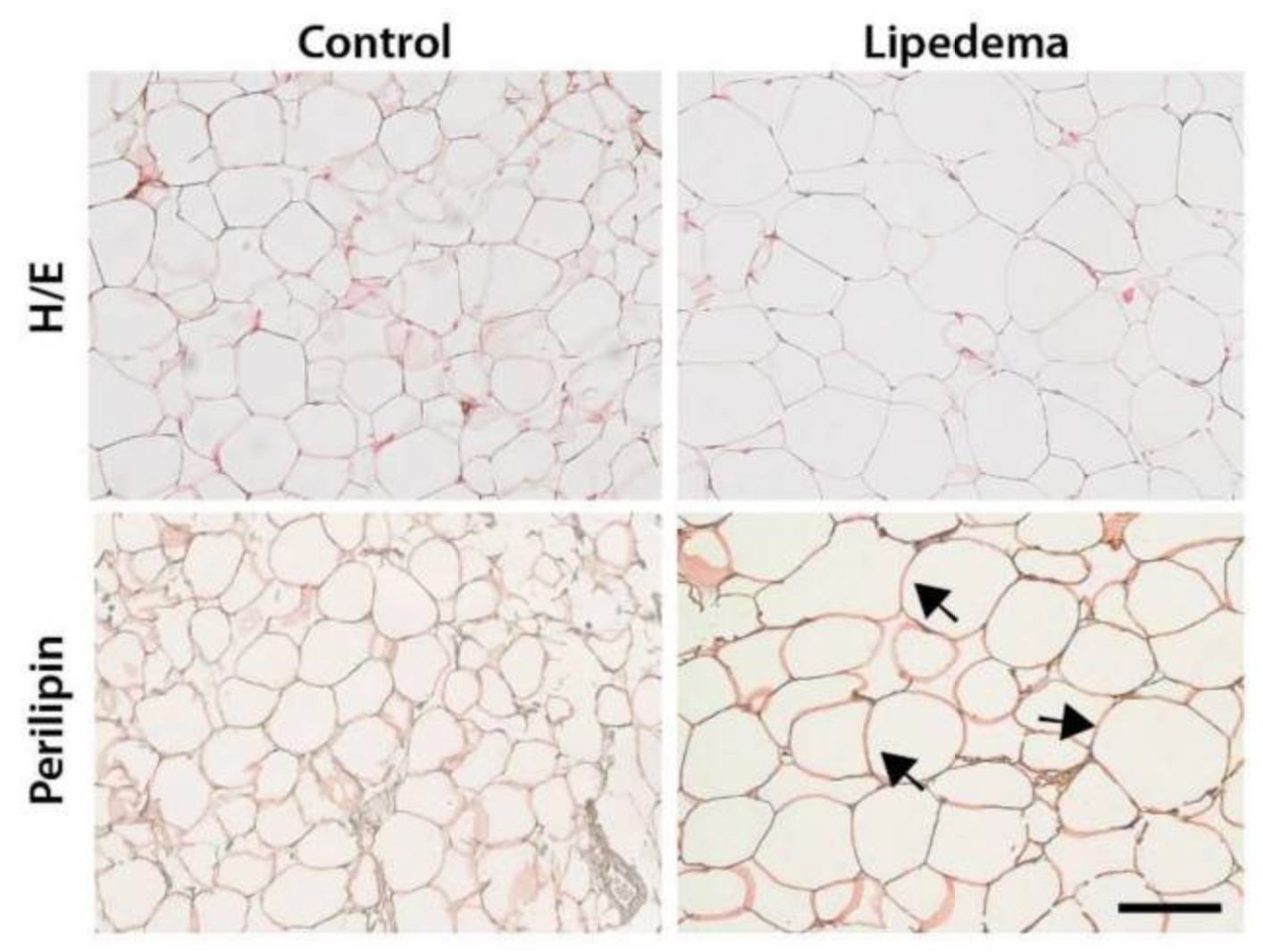
The researchers saw larger fat cells in lipedema tissue and a different pattern of immune signals (cytokines) in the blood compared with matched controls.

The word “cytokines” is something most women with lipedema have never heard of. As a food scientist, I’ve worked with cytokines in the laboratory, and I want you—who live with lipedema—to understand them too. If you have lipedema, you know the feeling: your legs, and sometimes your arms, behave as if they follow a different set of rules. They hurt, they bruise, they swell, and they don’t respond to weight loss the way the rest of the body does. And yet, when we try to prove that something real is happening under the surface, medicine still doesn’t have a simple blood test to point to.
Where and when this study was done
That’s why I want to show you this study from a serious clinical research environment: University Hospital Zurich in Switzerland, together with collaborators in Göttingen (Germany) and Turku (Finland). It was published in the International Journal of Molecular Sciences on March 24, 2021. The title is a mouthful, but the core question is simple and highly relevant: if we compare women with lipedema to women with the same body size (similar BMI) and similar age, can we find a biological “fingerprint” that separates lipedema from normal fat tissue—and maybe even points toward future biomarkers?
Why I read it differently
I’m reading this both as a curious lipedema patient and as a food scientist who has spent time studying cytokines and immune-modulating responses in human cells. And I can tell you: when researchers go beyond the scale and into tissue signals, things start to get interesting.
What the researchers wanted to explore
In this study, the researchers focused on three main areas. First, they looked at the fat cells themselves—are they different in size? Second, they examined the “fat chemistry,” meaning the types of fat molecules stored in tissue and circulating in blood. Third, they measured immune signaling in the blood and the metabolic “energy behavior” of the non-fat cells living inside fat tissue.
Who was included
They included 30 women in total: 20 with lipedema and 10 controls. The groups were matched so that BMI and age were comparable (average age in the late 40s, BMI around 27–28). The lipedema group included stages I to III, mostly stage II and III. Tissue samples were taken from the upper thigh, so the comparison was made from the same body area in both groups. In other words, they tried hard to compare like with like, so differences wouldn’t simply be explained by body weight.