
What Happens to Lipedema Tissue After Liposuction
A science-based look at tissue healing, long-term effects, and why liposuction reshapes lipedema symptoms without curing the disease.

When lipedema fat is removed through liposuction, the body does not simply fill an empty space. It initiates a coordinated healing program where the remaining tissue is cleared, repaired, and reorganized. Over time, this can shift the local environment away from the most pathologic features of lipedema tissue such as hypertrophied fat cells, inflammatory signaling, excess interstitial fluid, and fibrosis. The degree of lasting improvement varies, but the biology of healing helps explain why many patients experience durable symptom relief.
LipedemaScience was founded by a woman living with lipedema, diagnosed in 2012, who for many years experienced the lack of accessible and in-depth knowledge about the condition. Much has changed since then, and today an increasing amount of exciting research on lipedema is being published. She believes this knowledge should be available to everyone.
All articles are written by CarinaW, who has a background in laboratory research working with cells and DNA. You can support her work and help make research-based knowledge more accessible by becoming a member.
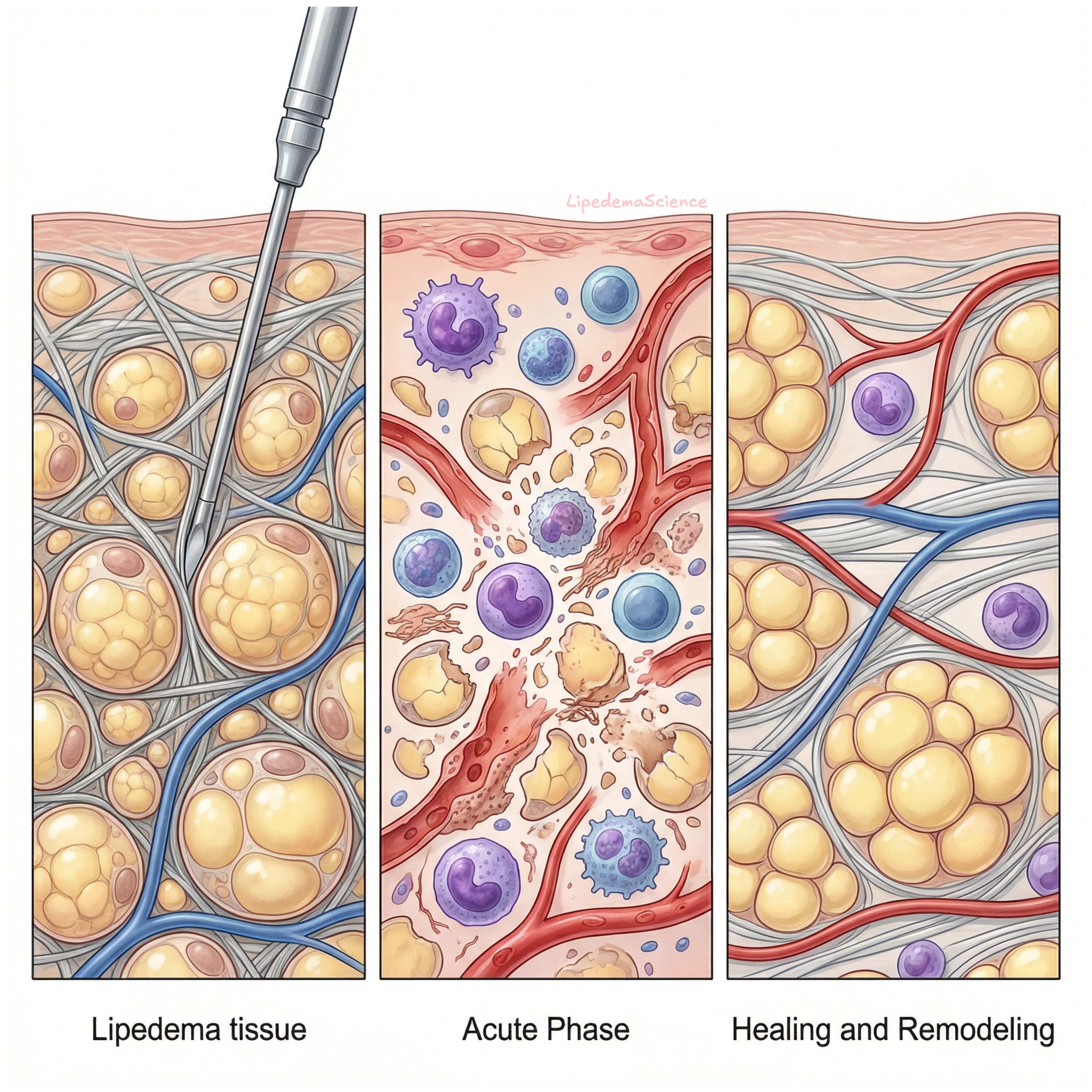
The procedure and the first trigger
Most lipedema surgery uses tumescent liposuction techniques designed to remove pathologic adipose tissue while aiming to limit harm to lymphatic structures. Even with careful technique, the procedure creates countless micro injuries in the surrounding connective tissue. Collagen fibers and the extracellular matrix are mechanically disrupted, small vessels are affected, and the body interprets this as an acute injury that requires an immediate inflammatory response. This early inflammatory step is not inherently negative. It is the signal that starts cleanup and rebuilding.
The acute phase clearing fluid and debris
Within hours to days, innate immune cells are recruited. Neutrophils arrive early, and macrophages follow to remove damaged cells and lipid remnants through phagocytosis. During this phase, cytokines and growth factors shape the next steps of repair. In parallel, the microvascular and lymphatic environment is actively remodeled. Signals that support new vessel formation and lymphatic adaptation can improve local drainage capacity and reduce the tendency toward persistent swelling. This is one reason why postoperative management that supports fluid movement can matter so much in the early weeks.
