I have spent a lot of time thinking about the term anti-inflammatory food. Not because I believe the concept is meaningless, but because I think we often use it with far more confidence than the underlying science allows.
This is particularly relevant in lipedema. Inflammation is discussed constantly in our community, and food is often presented as one of the main tools for controlling it. Certain foods are recommended because they are considered anti-inflammatory, others are removed because they are thought to promote inflammation, and entire dietary approaches are sometimes justified by the idea that they will reduce the inflammatory processes involved in lipedema.
I understand why this is appealing. I have used food as an important part of my own lipedema management for many years. I also have a Bachelor’s degree in Human Nutrition and a Master’s degree in Food Science, so food is not something I only approach from personal experience. During my MSc I worked with human intestinal cells, bioactive peptides derived from food proteins, inflammatory signalling and intestinal barrier function. Later in my laboratory career I also worked with bacterial DNA and whole-genome sequencing.
What my scientific education gave me was not a list of foods that are good or bad for inflammation. It gave me a much more complicated view of what happens between eating something and producing a biological effect.
My master’s thesis is a surprisingly useful example.
The experiment that could easily have become an “anti-inflammatory food” headline
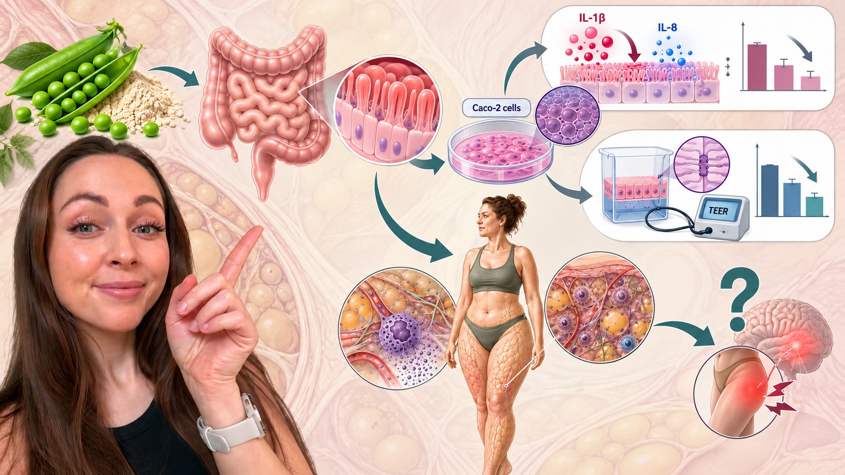
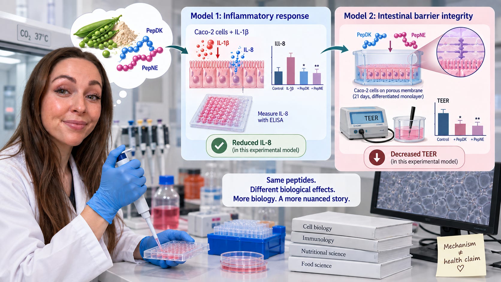
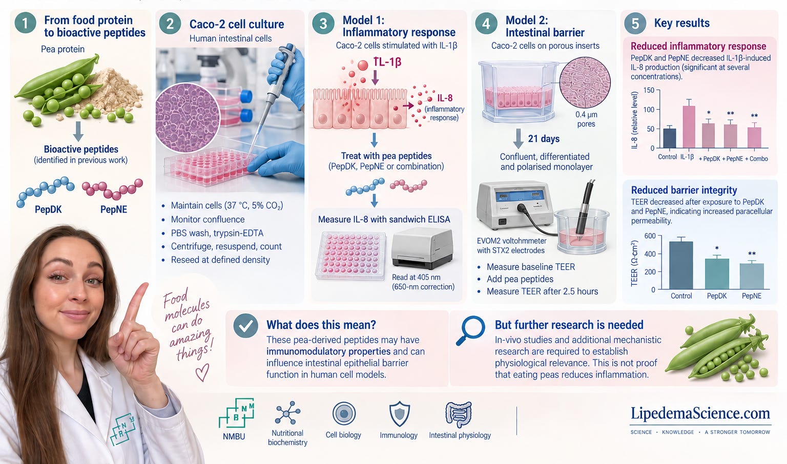
For my MSc in Food Science at the Norwegian University of Life Sciences, I investigated two peptides derived from pea protein. The peptides had already been identified in previous work within the research project. My part was to take those candidate molecules into biological models and ask whether they actually did anything to human intestinal cells.
I worked with Caco-2 cells, a human cell line widely used as an experimental model of intestinal epithelial cells. This involved ordinary mammalian cell culture work: maintaining the cells under controlled conditions, monitoring their growth, detaching and centrifuging them, counting them and reseeding them at defined densities. Anyone who has worked with cell culture knows how dependent the experiment is on the condition of the cells and consistency between experiments.
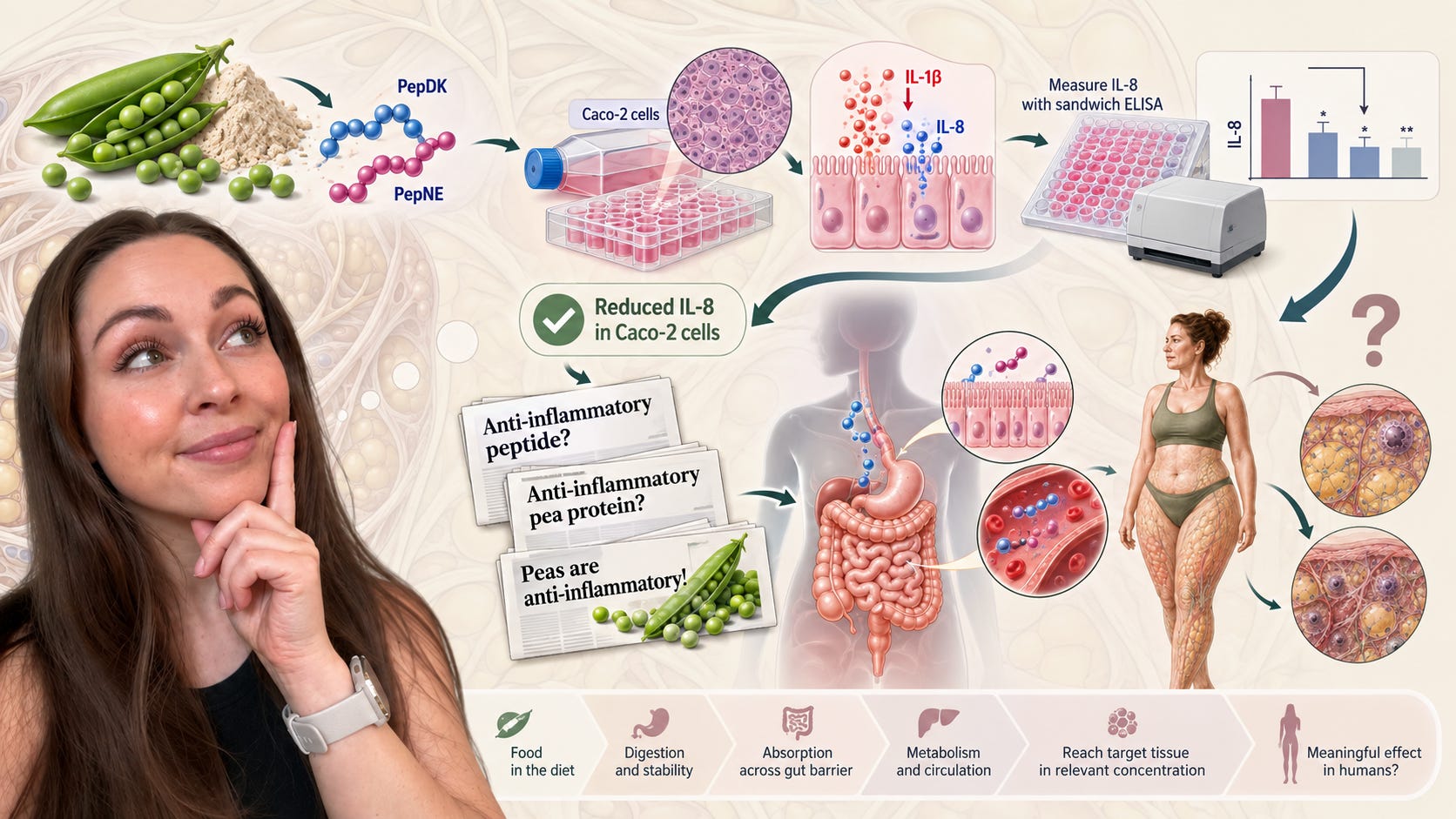
In one of my models, I deliberately provoked an inflammatory response. The cells were stimulated with IL-1β, an inflammatory cytokine, which resulted in the production of IL-8. I then exposed the cells to two pea-derived peptides, PepDK and PepNE, separately, together and at different concentrations. I quantified the amount of IL-8 using sandwich ELISA.
Several treatments reduced IL-1β-induced IL-8 production.
If I stopped describing the experiment at that point, I could construct a very attractive nutrition story from it. Pea-derived peptides reduced an inflammatory signalling molecule in human intestinal cells. It would not even be factually wrong to say that the peptides showed an anti-inflammatory or, more cautiously, an immunomodulatory effect in that experimental system.
The problem starts when the description of the experiment disappears but the adjective remains.
“Reduced IL-8 in IL-1β-stimulated Caco-2 cells under these experimental conditions” gradually becomes “anti-inflammatory peptide.” From there it can become “anti-inflammatory pea protein”, and eventually “peas are anti-inflammatory.”
Those statements do not carry the same amount of evidence.
This is one of the reasons I have become cautious about the language we use around anti-inflammatory food. A mechanistic experiment can tell us that a molecule is capable of interacting with a biological pathway. That is important information. But it does not tell us that eating the food containing that molecule will produce the same response in a human being, let alone in a particular tissue affected by a particular disease.
There are several steps in between. The compound has to exist in the food in a relevant amount. It has to survive food processing and digestion or be generated during digestion. It has to reach the relevant cells in a sufficient concentration. If we are claiming a systemic effect, it may have to cross the intestinal barrier, survive further metabolism, enter the circulation and reach another tissue. And then the effect seen in an isolated cellular system must still matter inside a human organism containing thousands of interacting signals, hormones, immune cells, metabolites and feedback systems.
That is the distance between mechanistic plausibility and clinical evidence.
What made my own results even more interesting
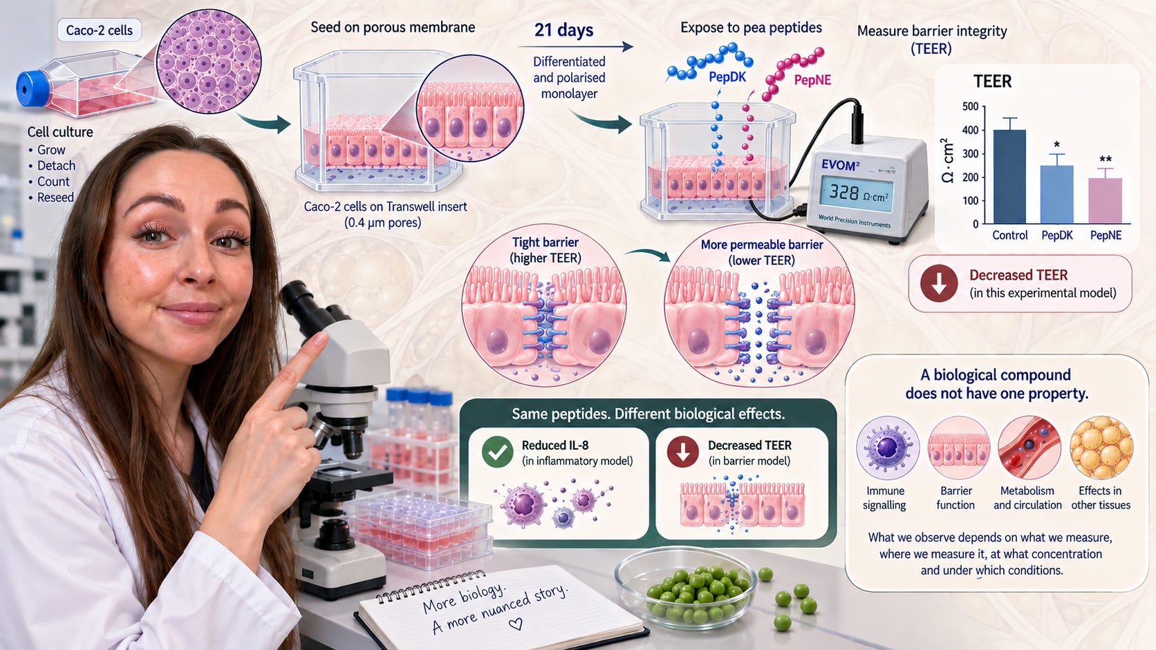
My thesis included a second Caco-2 model. Instead of looking primarily at inflammatory signalling, I grew the cells on porous membranes for approximately 21 days until they formed a differentiated and polarised epithelial monolayer, essentially a simplified model of the intestinal barrier.
I measured the integrity of that barrier using transepithelial electrical resistance, TEER. Higher resistance generally reflects a tighter epithelial layer, while falling resistance can indicate increased paracellular permeability.
When the mature cell layers were exposed to PepDK and PepNE, TEER decreased.
So the same peptides that could be described as “anti-inflammatory” based on the IL-8 experiment also altered another biological endpoint in a direction that, in the context of barrier integrity, would not automatically be interpreted as desirable.
That does not mean the peptides were harmful. The experiment was not designed to establish that, and a temporary change in TEER in an in-vitro model cannot be translated directly into a clinical judgement.
But it does demonstrate something that I think is frequently lost in nutrition discussions: a biological compound does not have one property.
A peptide is not simply anti-inflammatory. A fatty acid is not simply inflammatory. A polyphenol is not simply an antioxidant. These molecules enter biological systems containing receptors, enzymes, membranes and signalling networks. What we observe depends on what we measure, where we measure it, at what concentration and under which conditions.
If I had measured only IL-8, I would have had one story. Because I also measured epithelial resistance, I had a more complicated story.
The more biology you measure, the harder it often becomes to reduce a food to one adjective.
This becomes important when we move from food science to lipedema
The same problem appears in the current lipedema literature.
We increasingly have evidence that lipedema-affected adipose tissue is biologically interesting. Cifarelli and colleagues, for example, compared abdominal and thigh subcutaneous adipose tissue in women with obesity and lipedema. Within the women with lipedema, thigh tissue had more macrophages and greater expression of genes related to inflammation and fibrosis than abdominal adipose tissue, while expression of genes related to lymphatic and vascular biology was lower.
This is valuable because it examines the tissue that is actually affected rather than relying solely on blood markers.
But it also creates an important distinction. When we say that a diet “reduces inflammation,” what inflammation are we talking about?
CRP in blood?
IL-6?
TNF-α?
A change in circulating immune cells?
Macrophages inside thigh adipose tissue?
Expression of fibrosis-related genes?
Extracellular matrix remodelling?
Pain?
These outcomes are related to one another in some contexts, but they are not interchangeable.
The idea that lipedema is an inflammatory condition is often followed almost automatically by the conclusion that an anti-inflammatory diet should treat it. There is a missing step in that reasoning. In fact, there are several missing steps.
To make that mechanism convincing, we would ideally need evidence showing that a dietary intervention changes a relevant inflammatory pathway, that this change occurs in or meaningfully affects lipedema tissue, and that the biological change is associated with an improvement in clinically relevant outcomes such as pain or function.
We do not yet have that chain.
The low-carbohydrate trial is a good example of why this distinction matters
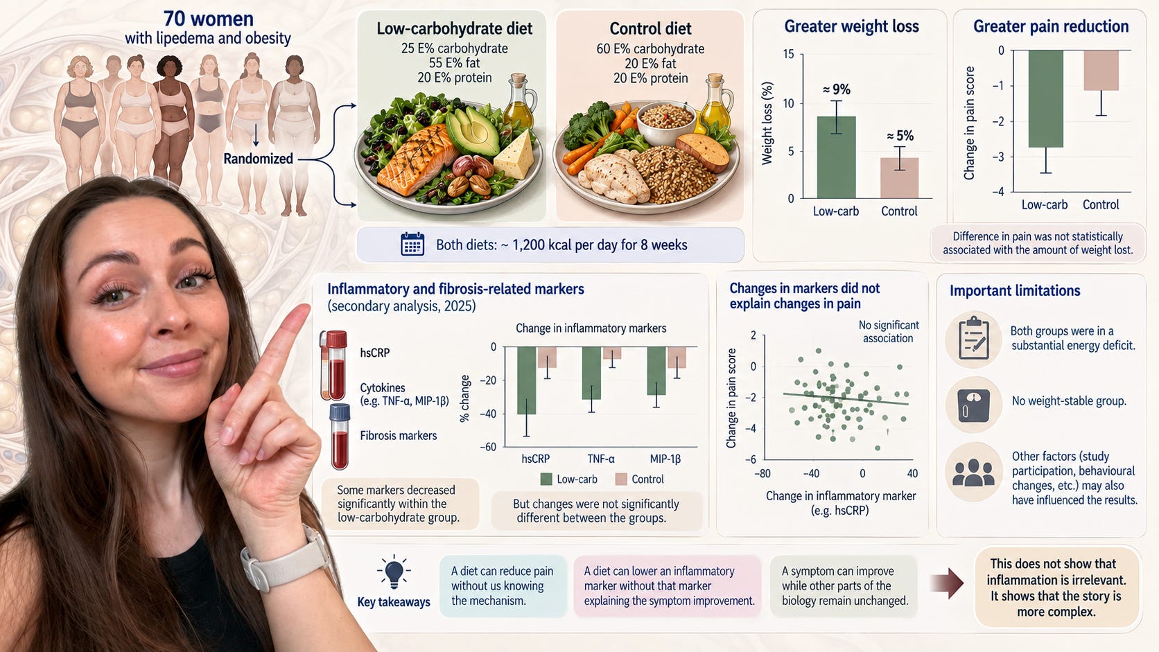
One of the most important dietary studies in lipedema so far is the Norwegian randomized controlled trial published in 2024. Seventy women with lipedema and obesity were randomized to either a low-carbohydrate diet or a control diet for eight weeks. Both diets were energy restricted to approximately 1,200 kcal per day. The low-carbohydrate group lost more weight and experienced a greater reduction in current pain than the control group. Importantly, the difference in pain was not statistically associated with the amount of weight lost.
That is genuinely interesting. It gives us considerably stronger evidence than anecdotes that dietary composition may matter for lipedema symptoms, at least in women with lipedema and obesity over an eight-week period.
But the next question is obvious: why did pain improve more?
A common explanation would be that the low-carbohydrate diet reduced inflammation.
Fortunately, the researchers actually investigated this rather than assuming it.
In a secondary analysis published in 2025, they measured hsCRP, multiple cytokines and fibrosis-associated markers. Some inflammatory markers, including TNF-α, macrophage inflammatory protein-1β and hsCRP, decreased significantly within the low-carbohydrate group. At first glance, that sounds like support for the anti-inflammatory explanation.
The between-group comparison is more important, however. The changes were not significantly different between the low-carbohydrate and low-fat groups. More importantly, changes in the measured cytokines and fibrosis-associated markers were not associated with changes in pain.
That does not show that inflammation has nothing to do with lipedema pain. It tells us something narrower and much more useful: the inflammatory markers measured in this trial did not explain why the low-carbohydrate group experienced greater pain reduction.
That is exactly the distinction I wish we made more often.
A diet can reduce pain without us knowing the mechanism.
A diet can lower an inflammatory marker without that marker explaining the symptom improvement.
And a symptom can improve while other parts of the biology remain relatively unchanged.
Those are not disappointing results. They are how science becomes more precise.
There is another limitation worth remembering. Both groups were placed on a substantial energy deficit. There was no weight-stable group eating the same diets. We therefore cannot cleanly separate everything caused by dietary composition from everything caused by negative energy balance, weight loss, study participation and other changes that accompany a structured intervention.
This does not invalidate the trial. It simply defines what the trial can and cannot answer.
What happens when we look directly at lipedema adipose tissue after weight loss?
This is where the Cifarelli study becomes particularly relevant.
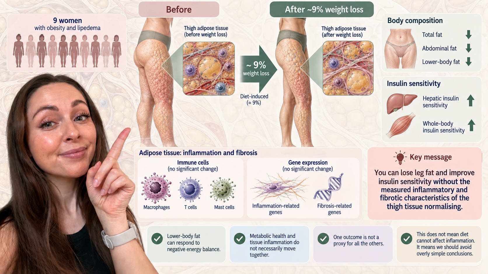
Nine women with obesity and lipedema were studied again after approximately 9% diet-induced weight loss. The researchers did not only measure their body weight. They measured insulin sensitivity, body-fat distribution and adipose-tissue biology.
The women lost total fat, abdominal fat and lower-body fat. Hepatic insulin sensitivity increased substantially and whole-body insulin sensitivity also improved. The common statement that lower-body fat in lipedema cannot respond to negative energy balance was therefore not supported by these data.
But the tissue results were striking for another reason.
Despite the metabolic improvements and loss of thigh fat, weight loss did not significantly alter adipose-tissue immune-cell content or the expression of genes associated with inflammation and fibrosis in the abdominal or thigh tissue.
This is extremely useful when thinking about “anti-inflammatory nutrition.”
It tells us that body composition, insulin sensitivity and local adipose-tissue inflammatory biology do not necessarily move together.
Someone can lose leg fat and improve insulin sensitivity without the measured inflammatory and fibrotic characteristics of the thigh tissue normalising over the same period.
Again, this does not mean diet cannot affect inflammation in lipedema. The sample was very small, the weight-loss period was relatively short, and there may be inflammatory processes the study did not measure.
It means we should stop assuming that one outcome is a proxy for all the others.
What about dietary inflammatory scores?
There is now also observational work relating diet quality to circulating inflammation in lipedema.
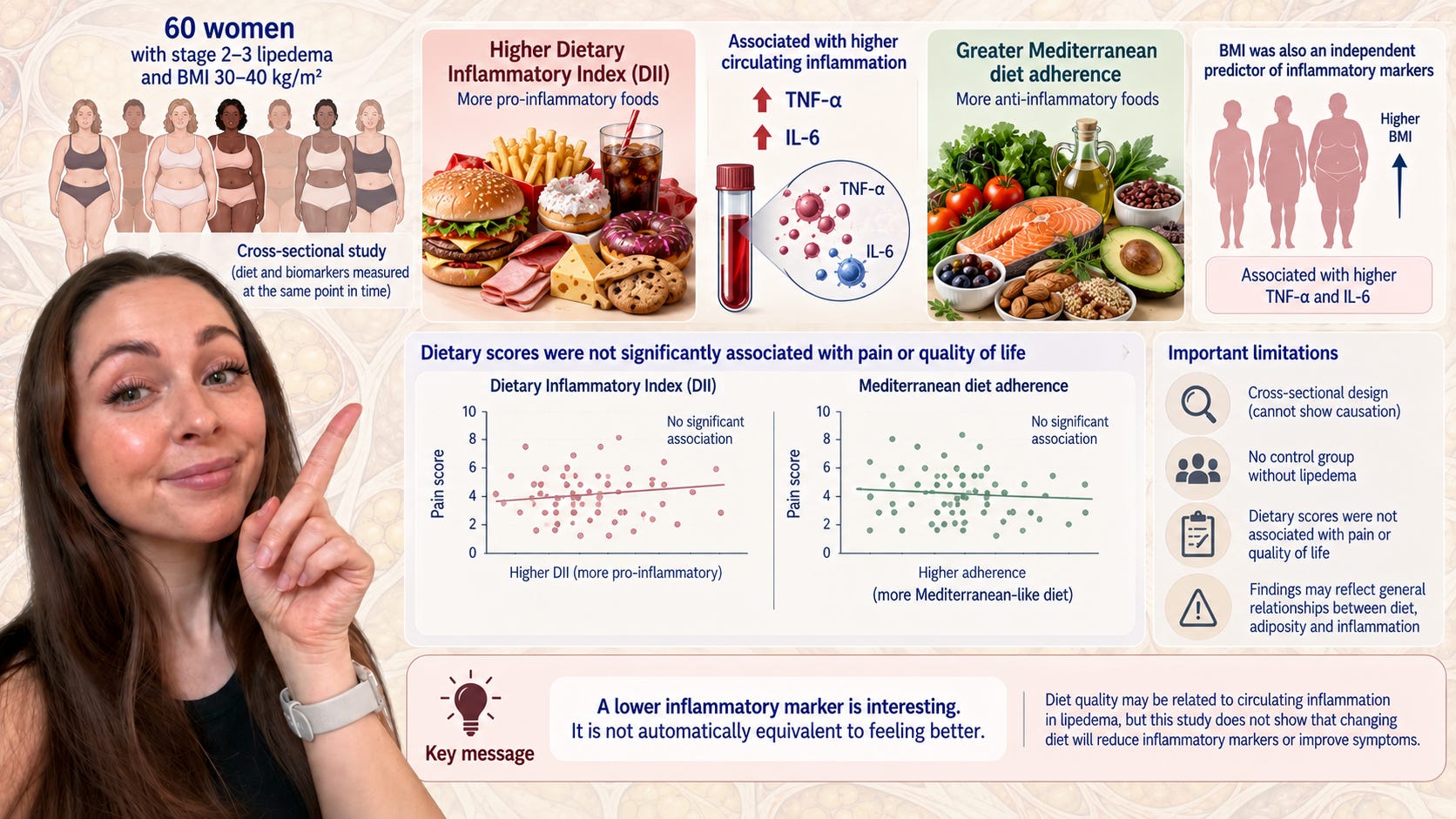
A study of 60 women with stage 2 or 3 lipedema and BMI between 30 and 40 kg/m² examined the Dietary Inflammatory Index and adherence to a Mediterranean dietary pattern. A higher Dietary Inflammatory Index was associated with higher circulating TNF-α and IL-6, while greater Mediterranean diet adherence was associated with lower levels. BMI was also an independent predictor of the inflammatory markers.
This is worth studying, but the interpretation has to match the design.
It was cross-sectional. The researchers looked at diet and biomarkers at the same point in time. They did not randomly change participants’ diets and demonstrate that doing so caused TNF-α or IL-6 to fall.
There was also no control group without lipedema, which means the study cannot establish that these relationships are specific to lipedema rather than reflecting relationships between dietary pattern, adiposity and systemic inflammatory markers that are also seen more broadly.
And importantly, the dietary scores were not significantly associated with pain or quality of life.
That last point is easy to overlook because the biomarker finding sounds more scientific. But from the perspective of somebody living with painful lipedema, it matters enormously.
A lower inflammatory marker is interesting. It is not automatically equivalent to feeling better.
Blood is not thigh adipose tissue
This may be the most important issue in the entire discussion.
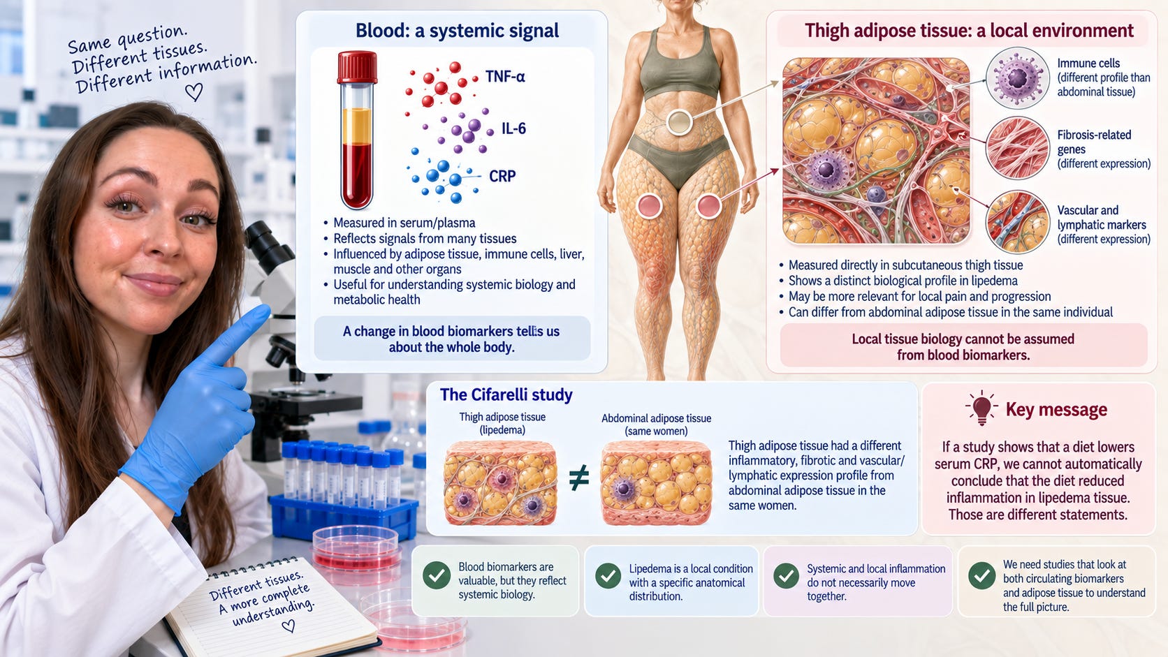
When we measure TNF-α, IL-6 or CRP in serum, we are measuring a systemic signal. Those concentrations are influenced by many tissues and many aspects of physiology.
Lipedema, however, is characterised by a very specific anatomical distribution. If the biological processes relevant to pain and progression are occurring locally in subcutaneous thigh tissue, circulating biomarkers may provide only a partial view.
The Cifarelli study makes this especially clear because thigh adipose tissue had a different inflammatory, fibrotic and vascular/lymphatic expression profile from abdominal adipose tissue in the same women.
This does not mean blood biomarkers are useless. They can tell us about systemic biology, and systemic metabolic health is important regardless of lipedema.
But if a study shows that a diet lowers serum CRP, we cannot automatically rewrite that result as “the diet reduced inflammation in lipedema tissue.”
Those are different statements.
The intestinal barrier is another area where I think we need restraint
Because I worked with an intestinal barrier model during my MSc, I find the current interest in gut permeability particularly interesting.
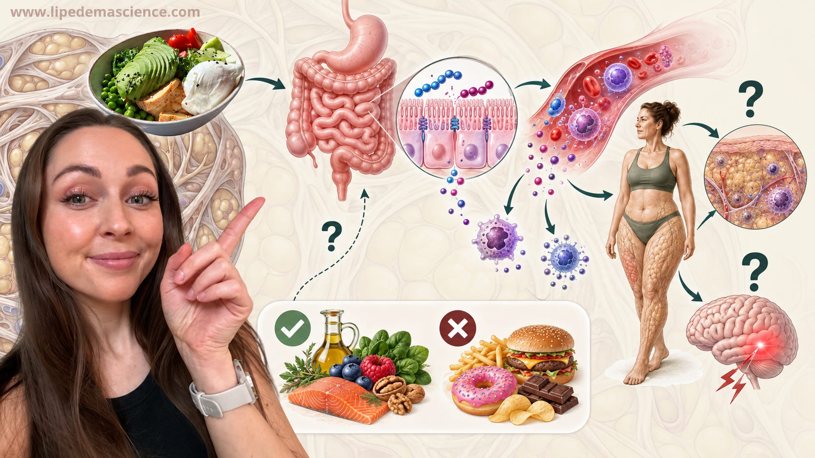
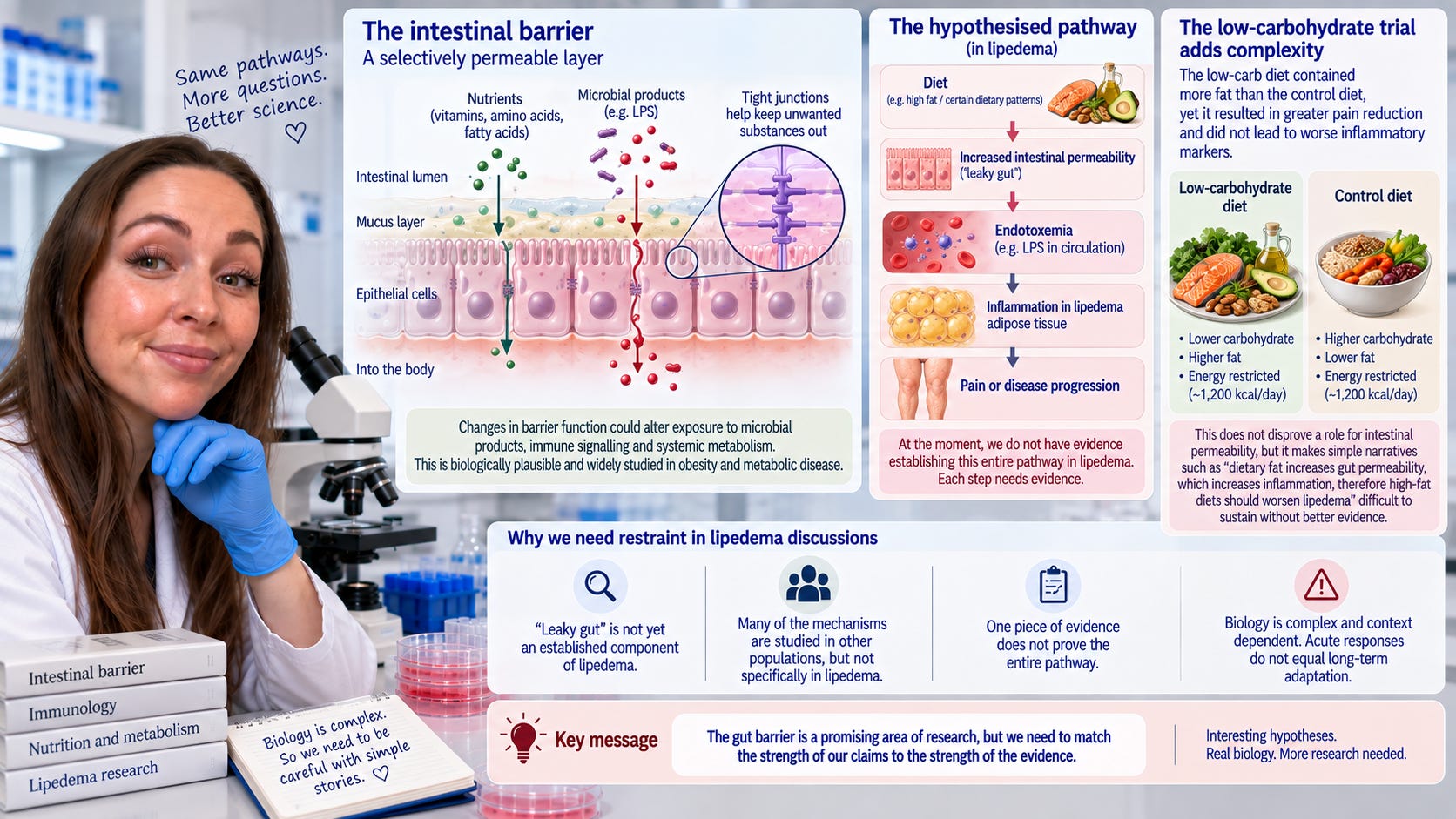
The general hypothesis is biologically plausible. Changes in intestinal barrier function could alter exposure to microbial products, immune signalling and systemic metabolism. There is a large research literature on these mechanisms in obesity and metabolic disease.
But I frequently see this taken much further in lipedema discussions, where “leaky gut” is presented almost as an established component of the disease.
At the moment, we do not have evidence establishing the following pathway in lipedema:
diet → increased intestinal permeability → endotoxemia → inflammation in lipedema adipose tissue → pain or disease progression.
Each arrow needs evidence. The existence of research supporting one arrow in another population does not establish the entire pathway in women with lipedema.
There is even an interesting tension in the available dietary research. The low-carbohydrate intervention in the Norwegian trial contained considerably more fat than the comparison diet, yet it produced greater pain reduction and did not result in significantly worse changes in the measured inflammatory markers.
That does not disprove a role for intestinal permeability. Acute responses to a high-fat meal are not equivalent to adaptation to a dietary pattern over eight weeks, and the quality of fat, energy balance, weight loss and many other variables could alter the response.
But it does make a simple story such as “dietary fat increases gut permeability, which increases inflammation, therefore high-fat diets should worsen lipedema” difficult to sustain without much better evidence.
Biology rarely gives us such clean narratives.
So is an anti-inflammatory diet useless?
No. That is not the conclusion I draw from any of this.
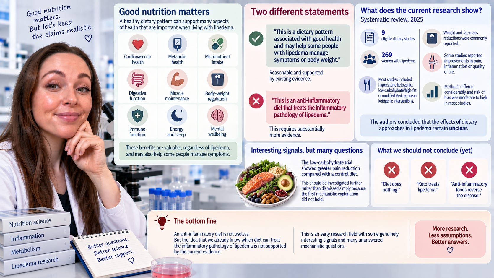
I think good nutrition matters enormously, including for people with lipedema. A dietary pattern can support cardiovascular health, metabolic health, adequate micronutrient intake, digestive function, muscle maintenance, body-weight regulation and many other things that matter when living with a chronic condition.
There may also be dietary approaches that affect lipedema symptoms through mechanisms we do not yet understand. The low-carbohydrate pain data are a good example. That finding should be investigated further rather than dismissed simply because the first mechanistic explanation did not hold.
But we should distinguish between two very different statements:
“This is a dietary pattern associated with good health and may help some people with lipedema manage symptoms or body weight.”
and
“This is an anti-inflammatory diet that treats the inflammatory pathology of lipedema.”
The second statement requires substantially more evidence.
A systematic review published in 2025 found only nine eligible dietary studies involving 269 women with lipedema. Most involved hypocaloric ketogenic, low-carbohydrate/high-fat or modified Mediterranean ketogenic interventions. Weight and fat-mass reductions were commonly reported, and some studies reported improvements in pain, inflammation or quality of life, but methods differed considerably and risk of bias was moderate to high in most studies. The authors concluded that the effects of dietary approaches in lipedema remain unclear.
That is the current state of the evidence.
It is not “diet does nothing.”
It is not “keto treats lipedema.”
It is not “anti-inflammatory foods reverse the disease.”
It is an early research field with some genuinely interesting signals and many unanswered mechanistic questions.
My MSc changed the way I think about food, but probably not in the way people expect
When I studied Human Nutrition, I learned a great deal about energy, nutrients, metabolism, requirements and health.
Food Science took me deeper into what food actually is biologically.
A protein is not only protein. Digestion produces peptides and amino acids. Plant foods contain compounds capable of interacting with enzymes and receptors. Fatty acids can participate in signalling. Food components can affect cellular responses, gene expression and barrier properties.
This is why I often say that food is much more than macros.
But there is another half to that statement that I think is just as important.
Because food is biologically complex, we should be less willing to classify foods according to one simple biological effect.
My own master’s thesis showed me this very clearly. The same pea-derived peptides reduced IL-8 production in one experiment and reduced TEER in another. The honest conclusion was not that peas were anti-inflammatory, nor that they were harmful to the gut. The honest conclusion was that these peptides produced measurable biological effects in two Caco-2 models, and that further research was needed to understand their physiological relevance.
That may sound less exciting than a headline promising anti-inflammatory food.
For me, it is much more interesting.
It forces us to ask the questions that matter. What exactly was measured? Was it measured in a cell, in blood or in the affected tissue? Was the study observational or experimental? Did a biomarker change? Did symptoms change? Did the biomarker explain the symptoms? Was there a control group? Was the effect independent of weight loss or energy restriction? And, perhaps most importantly, how many assumptions are we making between the result of the experiment and the advice we give to a person with lipedema?
Those are the questions I want to bring into my writing about nutrition.
Not because I think food is unimportant in lipedema. Quite the opposite. I think food is biologically fascinating and potentially very important. But I do not think people with lipedema benefit from being given another long list of foods they are told to fear because somebody once measured an inflammatory marker.
We already carry enough responsibility for managing this condition.
The research should help us understand our bodies better, not create a new form of guilt every time we sit down to eat.