

Yesterday, Amber K asked a great question in the LipedemaScience member chat! After reading my interview with Dr. Deise Vargas earlier this week, she wanted a simpler explanation of how ultrasound can identify differences in tissue. So, Amber, this one is for you!
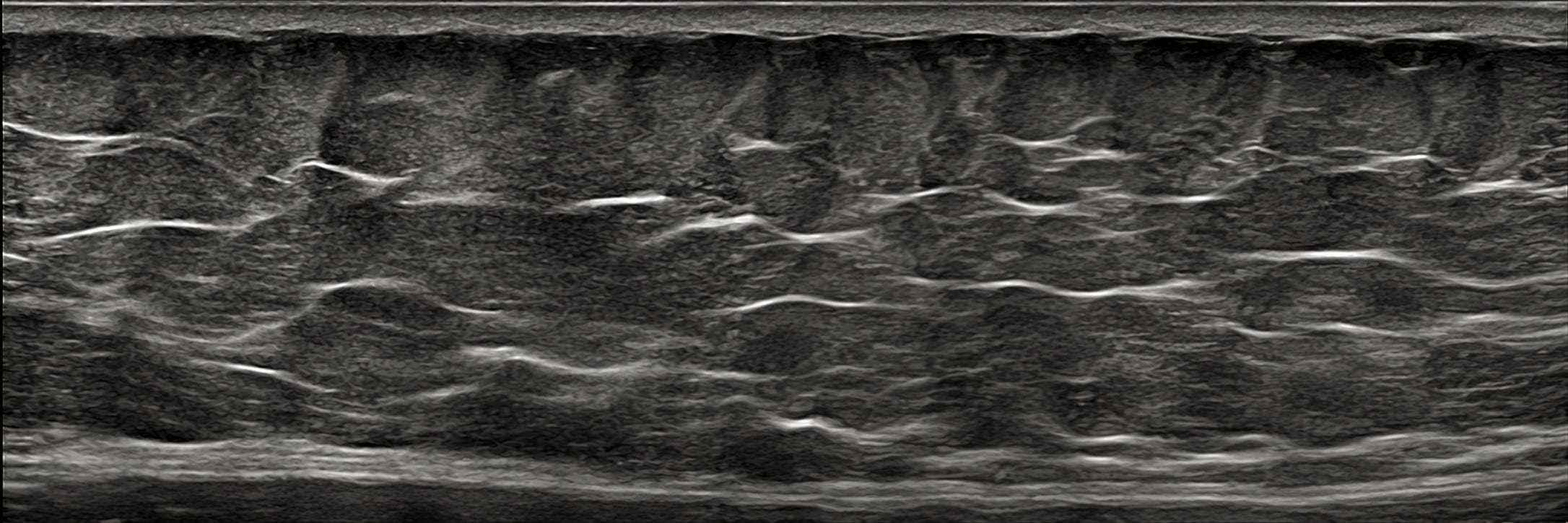
Think of ultrasound as a camera beneath your skin
Ultrasound sends sound waves into your body. These waves bounce back differently depending on what they encounter, such as fat, connective tissue, or muscle. A computer turns those echoes into an image.
But what does this have to do with lipedema?
Under your skin, fat is arranged in small groups called fat lobules, held together by connective tissue. Think of it like a bunch of grapes, where the grapes represent fat and the structures holding them together represent connective tissue.
In Dr. Vargas’s research, ultrasound revealed that this arrangement can look different in people with lipedema. Rather than simply measuring how much fat someone has, she examines how the tissue is organized.
Researchers can look for:
Changes in fat tissue: Whether the fat lobules appear differently arranged or the tissue looks thicker.
Changes in connective tissue: Whether the structures separating and supporting the fat appear altered or disrupted.
Small nodules: Localized areas that look different from the surrounding tissue. Some painful nodules investigated by Dr. Vargas contained evidence of small bleeding events and damaged fat cells.
