
The Silent Conversation Inside Fat Cells
How hormone receptors shape the biology of Lipedema. A deeper layer of communication.

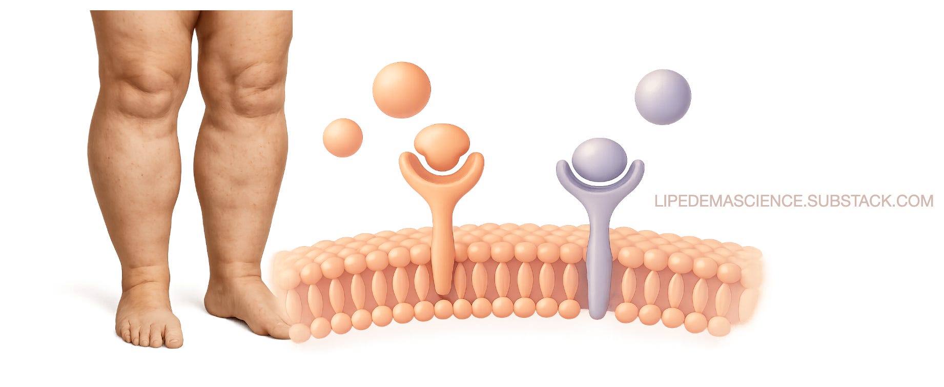
Inside every cell of the body, hormones act as messengers. But in lipedema, this cellular conversation seems to have changed its tone. Researchers are discovering that the receptors—the molecular “ears” that listen to hormones like estrogen and progesterone—behave differently in women with lipedema. This shift may be one of the keys to understanding why fat tissue in lipedema grows, hurts, and resists typical weight-loss strategies.
Three receptors, one delicate balance
Estrogen doesn’t act alone. Its message is carried through three receptors: ERα, ERβ, and GPER.
In healthy tissue, ERα supports metabolic health, insulin sensitivity, and normal fat distribution, while ERβ keeps growth and inflammation in check. GPER, a third receptor found in the cell membrane, helps regulate energy balance and pain signaling.
In lipedema, this harmony is lost. Studies show ERα is reduced and ERβ is elevated, creating a receptor imbalance that encourages inflammation, fibrosis, and impaired fat breakdown. GPER often decreases as well—another hit to the tissue’s ability to regulate metabolism and energy use.
When the signals go wrong
This receptor shift alters how the fat tissue behaves. The result is not just fat accumulation but a fundamental change in the tissue’s biology. Cells grow larger and stiffer, inflammatory molecules increase, and the fine network of tiny blood vessels becomes leaky and fragile. Over time, this promotes swelling, pain, and the thickened texture many women notice under the skin.
Laboratory studies support these findings. When fat-derived stem cells from women with lipedema are exposed to estrogen, they react abnormally—showing signs of enhanced fat-cell formation and altered gene activity compared to healthy controls. It’s as if the tissue interprets estrogen in a distorted way.
Fat that makes its own hormones
A fascinating aspect of lipedema biology is that the affected tissue produces its own estrogen. Enzymes such as aromatase, 17β-HSD1, and 17β-HSD7 convert weaker hormones into active estradiol, while enzymes like 17β-HSD2 and SULT1E1—which normally inactivate estrogen—are reduced. This creates a self-sustaining loop of estrogen activity inside the tissue, even when blood levels of the hormone are low, such as after menopause.
Researchers call this intracrine signaling—a local hormone system that operates independently of the rest of the body. In lipedema, this local estrogen production may drive ongoing inflammation, swelling, and tissue remodeling long after systemic hormones have declined.
Progesterone’s missing brake
Normally, progesterone acts as a counterbalance to estrogen. It calms inflammation, limits fibrosis, and keeps tissue repair in check. But evidence from related estrogen-driven conditions—such as endometriosis and uterine fibroids—suggests that progesterone resistance may develop in lipedema too. When the progesterone “brake” fails, estrogen’s effects dominate, and tissue inflammation accelerates.
Shared pathways with other women’s health disorders
The receptor pattern in lipedema mirrors what researchers have seen in other chronic gynecologic conditions: a shift toward ERβ dominance, local estrogen excess, and progesterone resistance. These shared mechanisms link lipedema not just to fat metabolism, but to a broader hormonal landscape that includes endometriosis, adenomyosis, and fibroids. Recognizing these common roots could eventually help clinicians adapt treatment strategies across conditions.
What this means for treatment
Understanding hormone receptors opens new perspectives, even if clinical applications are still evolving. Traditional care—compression, lymphatic therapy, physical activity, and, when needed, liposuction—remains essential. But as science advances, targeting receptor balance could complement these measures.
Future research may explore therapies that modulate ERα/ERβ activity, normalize local hormone metabolism, or restore progesterone responsiveness. For now, these insights help explain why lipedema behaves differently from obesity—and why treatment must go deeper than calorie counts.
Listening to the body’s language
Every discovery about hormone receptors brings us closer to seeing lipedema as a biologically complex, hormonally sensitive disorder rather than a cosmetic or lifestyle issue. The tissue itself is not passive—it listens, reacts, and sometimes misinterprets the body’s hormonal cues.
Understanding that silent conversation is the first step toward restoring balance—one receptor, one signal, and one woman at a time.
Management of Lipedema with Ketogenic Diet: 22-Month Follow-Up (DOI: 10.3390/life11121402)
Observational Study on a Large Italian Population with Lipedema: Biochemical and Hormonal Profile, Anatomical and Clinical Evaluation, Self-Reported History (DOI: 10.3390/ijms25031599)
Lipedema and the Potential Role of Estrogen in Excessive Adipose Tissue Accumulation (DOI: 10.3390/ijms222111720)
Lipedema: From Women’s Hormonal Changes to Nutritional Intervention (DOI: 10.3390/endocrines6020024)
Reproductive Landmarks and Lipedema: Lessons to be Learned about Women Hormones throughout Life (DOI: 10.5772/intechopen.1006956)
Estrogen as a Contributing Factor to the Development of Lipedema (DOI: 10.5772/intechopen.96402)
Menopause as a Critical Turning Point in Lipedema: The Estrogen Receptor Imbalance, Intracrine Estrogen, and Adipose Tissue Dysfunction Model (DOI: 10.3390/ijms26157074)
Lipedema as a Hormone-Driven Gynecological Disorder: The Estrogen Receptor Connection (DOI: 10.5772/intechopen.1012036)
The Expression of Adipogenic Marker Is Significantly Increased in Estrogen-Treated Lipedema Adipocytes Differentiated from Adipose Stem Cells In Vitro (DOI: 10.3390/biomedicines12051042)