

In a case series published in Cureus in August 2026, eight painful lumps were surgically removed from four women because doctors suspected angiolipoma, a small benign tumor of fat cells and tiny blood vessels that often sits tender under the skin. Two of the women had angiolipomas. The six lumps from the other two, who both had lipedema, turned out to be old bleeding inside the fat. The authors use this contrast to propose ultrasound criteria that could spare women with lipedema an unnecessary operation, and this article looks at what the findings support and what they do not yet support.
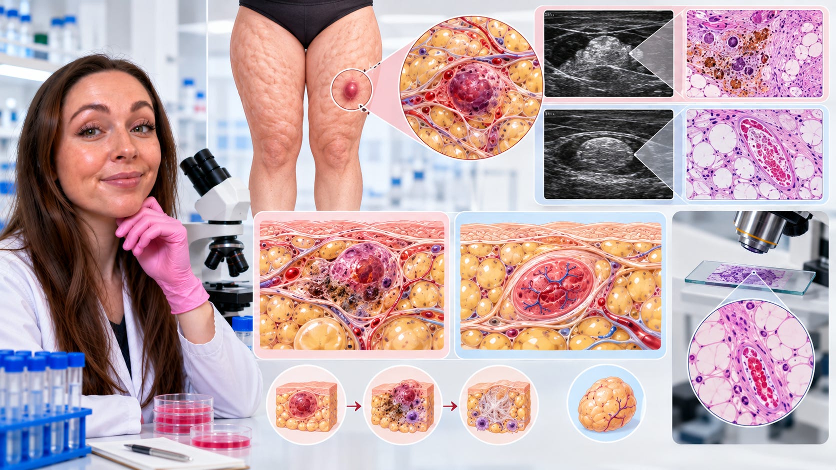
What was found
The two women with lipedema, aged 55 and 36, contributed six nodules, and the two without lipedema, aged 38 and 32, each had one lump on the thigh. All lumps were scanned using the Lipedema Dermal and Hypodermal Classification (LDHC), a qualitative ultrasound system developed by authors of this paper. Its third pattern includes nodules that appear brighter than the surrounding tissue, called hyperechoic, set in disorganized superficial fat with irregular connective tissue strands. Under the microscope, the lipedema nodules held leaked blood, hemosiderin, new fibrous tissue and patches of dead fat cells. Hemosiderin is the iron pigment left when red blood cells break down, so the bleeding was not fresh.
