

For many women with lipedema, pain, tenderness, swelling, and changes in the tissue beneath the skin are part of everyday life. Yet understanding what actually happens inside lipedema tissue, and why it can become so painful, remains one of the important unanswered questions in lipedema research.
Could ultrasound help us see changes that conventional examinations may overlook?
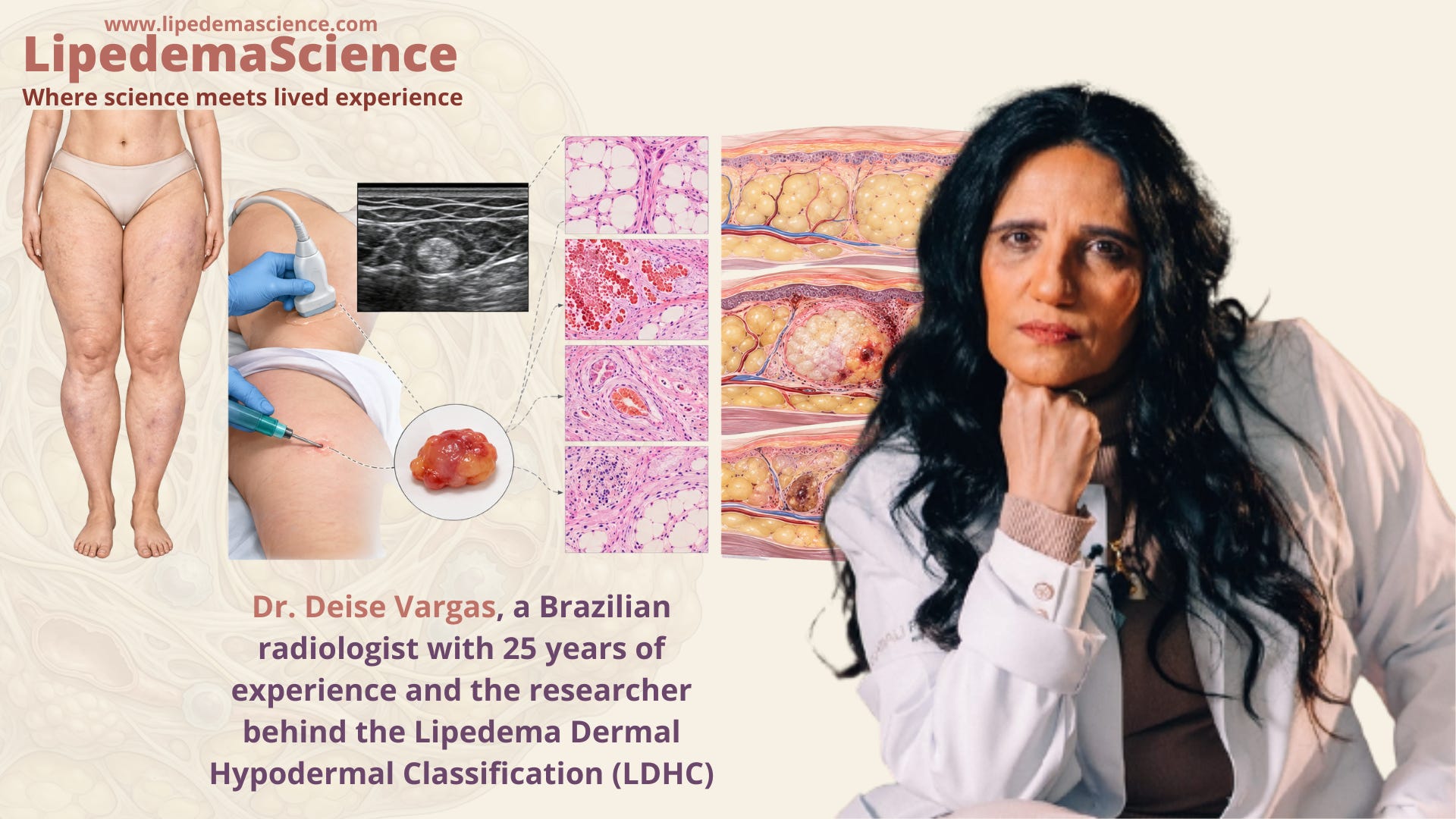
In this exclusive LipedemaScience interview, I speak with Dr. Deise Vargas, a Brazilian radiologist with 25 years of experience and the researcher behind the Lipedema Dermal Hypodermal Classification (LDHC). Her work explores how ultrasound imaging can reveal structural changes in the skin and subcutaneous adipose tissue, including alterations in tissue organization, fibrosis, and small, painful nodules.
Particularly interesting are her team’s investigations into microvascular changes, localized bleeding, fat necrosis, and possible mechanisms of lipedema pain. By combining ultrasound findings with tissue biopsies, the researchers are exploring whether some of these changes could help explain symptoms that have historically been difficult to assess objectively.
Throughout our conversation, Dr. Vargas explains what her research has revealed so far, why certain ultrasound findings should not be confused with angiolipomas, and how imaging might eventually contribute to monitoring lipedema progression and treatment outcomes. We also discuss the limitations of the current evidence, including why ultrasound cannot yet replace clinical diagnosis and why independent validation remains essential.
This interview offers a fascinating look at an emerging area of lipedema research, where the goal is not simply to measure how much adipose tissue is present, but to better understand what is happening within that tissue.

1 - Could you tell us about your background as a radiologist? What first drew your attention to lipedema?
I am a Brazilian radiologist with 25 years of experience, working primarily in dermatological, musculoskeletal, and rheumatological ultrasound, with a particular focus on lipedema.
I am a researcher and the creator of the ultrasound morphological classification of lipedema, the LDHC (Lipedema Dermal Hypodermal Classification), which describes four patterns of morphological alterations in lipedema in comparison with the normal pattern.
I have published scientific articles on international platforms and in international journals, including Cureus, Radiopaedia, and SCIRP. I am also the author of the books From Unlikely to Memorable: Discovering My Purpose in Lipedema and Lipedema in Layers. I have also been a speaker at several courses and congresses throughout Brazil on lipedema ultrasound.
I am the founder and Medical and Scientific Coordinator of LIPCOR (Lipedema and Diagnostic Correlations Center), dedicated to the study of lipedema, always with a focus on ultrasound correlation.
The beginning of my journey in lipedema did not take place in a laboratory, but rather through clinical observation. It began with the leg pain experienced by my patients, who were referred to me for vascular imaging evaluation. For more than two decades, while examining women referred for venous and arterial Doppler ultrasound, I repeatedly encountered the same situation: patients with severe pain, increased limb volume, and spontaneous bruising, yet whose vascular examinations were often normal. Nevertheless, these women continued to suffer and were sometimes even referred for varicose vein surgery.
In 2024, I began to recognize that there were alterations that conventional vascular imaging methods were failing to demonstrate. Then, in early 2025, one particular patient definitively changed the way I understood lipedema. She presented with clinical manifestations associated with profound suffering, which deeply affected me. Faced with her case, I began to question whether we were truly examining the tissue responsible for her symptoms. I realized that I needed to look beyond the vascular system and investigate the hypodermis.
That was when I began identifying ultrasound alterations in the organization of the hypodermis. When these findings were grouped together, they corresponded to morphological patterns that warranted systematic investigation.
From this observation, my journey as a lipedema researcher began, together with the purpose of developing an ultrasound methodology capable of documenting these alterations, reducing subjectivity in their assessment, and contributing to the monitoring and follow-up of these patients.
Therefore, my research was born from a very simple question:
“How can an examination be considered normal when the patient continues to experience so much pain?”
2 - Was there a particular patient, ultrasound finding, or unanswered question that led you to begin researching the tissue changes associated with lipedema?
Yes. She was exactly the patient I mentioned earlier. She represented a turning point in my scientific journey because it was during her examination that I began to understand that we had been evaluating patients in an incomplete way.
Until then, the ultrasound assessment of lipedema described in the literature had focused primarily on quantitative parameters, particularly the measurement of subcutaneous tissue thickness, an approach widely disseminated by Dr. Alexandre Amato, a Brazilian physician and researcher and a recognized reference in the field of lipedema.
However, in that patient, I realized that there was a need to understand the architecture of all layers of the dermis and hypodermis.
During the examination, I observed a constellation of morphological alterations that immediately caught my attention. I observed changes in tissue thickness, as previously described by Dr. Amato, but also alterations in the echogenicity of the hypodermis, associated with changes in the organization of the septa and fascial structures located between the adipose lobules.
I began to systematically assess five morphological aspects: tissue thickness, echogenicity, septal organization, alterations of the interfaces and fascial structures, and the presence of focal alterations in the architecture of the hypodermis and at the dermal–hypodermal junction.
As I began evaluating other patients with lipedema, I realized that these findings were not isolated observations. Rather, they represented recurring morphological patterns, although with different degrees of expression and tissue disorganization.
Within this context, I identified a “hyperechoic nodule” within the hypodermis. It was not clinically palpable, yet it was associated with marked pain upon digital pressure.
These observations led me to identify four distinct ultrasound patterns, which subsequently provided the foundation for the development of the LDHC (Lipedema Dermal Hypodermal Classification).
3 - You developed the LDHC ultrasound classification. What gap were you trying to address, and how would you explain the classification to someone without a medical background?
The research gap emerged from the absence of a standardized ultrasound methodology capable of identifying, characterizing, and classifying the morphological alterations of adipose tissue in patients with lipedema.
Imaging methods such as magnetic resonance imaging, computed tomography, and DEXA were already capable of providing important information about the distribution and quantity of adipose tissue. However, I was looking for something different: I wanted to understand and demonstrate the alterations in the tissue architecture itself through ultrasound, and to give the disease an imaging representation.
It was from this need that the LDHC (Lipedema Dermal Hypodermal Classification) was developed.
The proposal was to establish morphological criteria that would allow the assessment of alterations from the skin surface through the different compartments of the hypodermis. Measuring hypodermal thickness as a diagnostic parameter may produce false-positive results, for example, in patients with obesity or edema. The distinction is that we do not want to know only how much adipose tissue is present, but rather how that tissue is organized.
The LDHC also opens the possibility of monitoring these patients, including their tissue alterations, clinical evolution, and responses to different treatments.
It is important to clarify that the LDHC was developed as a complementary morphological classification. Its diagnostic application and prognostic value still require validation in larger populations and by independent research groups.
My purpose was to provide lipedema with an ultrasound representation of its tissue architecture, so that we could not only measure the tissue, but also begin to understand and objectively document its alterations.
4 - What first made you suspect that some painful nodules in lipedema might be mistaken for angiolipomas?
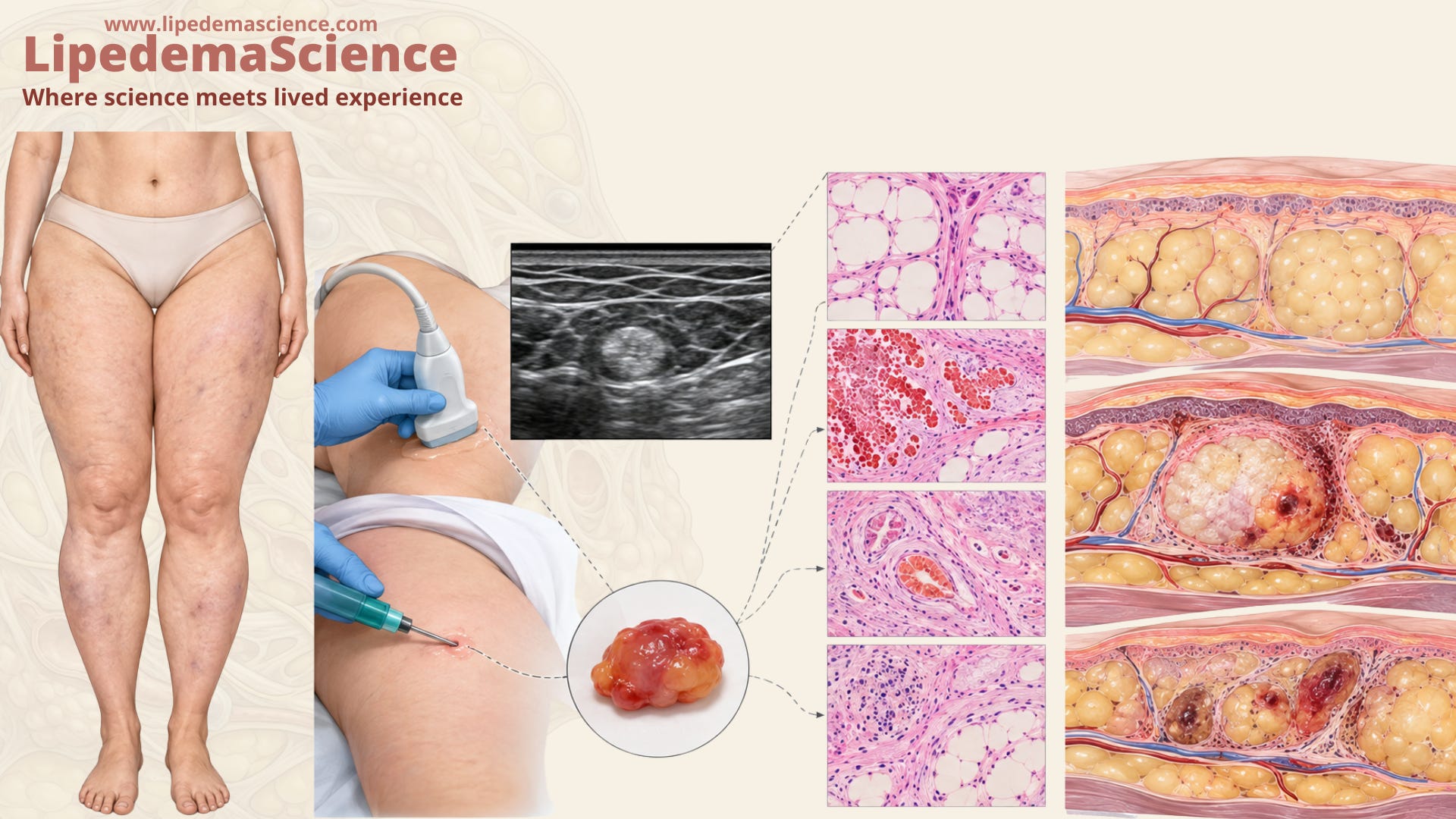
During the development of the LDHC classification, hyperechoic nodules were observed exclusively in LDHC Type 3. These nodules are small, generally less than 1 cm in diameter, not clinically palpable, and markedly painful. They are distributed within adipose tissue that already exhibits structural alterations.
To investigate the etiology of these nodules, we performed an excisional biopsy for histopathological analysis. Microscopy demonstrated that the nodules represented areas of hemorrhage associated with fat necrosis, hemosiderin deposition, and immature neoangiogenesis. These findings were interpreted as evidence of a hemorrhagic process occurring within a hypoxic–ischemic tissue environment.
It is essential to clarify that these nodules are not pathognomonic of lipedema. However, they are frequently observed in patients with lipedema.
Angiolipoma is a benign tumor composed of adipose tissue and a vascular component and may also present with pain. Therefore, these clinical characteristics could potentially lead to confusion between an angiolipoma and the LDHC Type 3 nodule.
Because I have extensive experience in dermatological ultrasound, using high-frequency, high-resolution transducers, I began to investigate the morphological characteristics of the LDHC Type 3 nodules in greater detail. I recognized that these nodules displayed characteristics that differed from those I typically observed in angiolipomas.
The most important insight was realizing that our objective was no longer simply to identify the hyperechoic nodule on ultrasound. Instead, we began to ask what this finding might reveal about the underlying processes occurring within the hypodermis of patients with lipedema.
This observation may represent a shift in the way we understand the pathophysiology of lipedema. Traditionally regarded primarily as an inflammatory condition, lipedema may also involve ischemic changes within the tissue, particularly in the context of the LDHC Type 3 pattern.
Thus, the hyperechoic nodule may be more than an isolated ultrasound finding. It may represent an imaging marker of underlying tissue injury and provide a window into the microvascular and hypoxic–ischemic processes occurring within the hypodermis.
5 - In your recent case series, what were the clearest differences between the nodules on ultrasound? What did the tissue samples add to your understanding?
The decision to perform biopsies arose from the need to differentiate structures that may exhibit similar ultrasound appearances when the nodules are analyzed in isolation, but that have different origins and require different therapeutic approaches.
The hyperechogenicity of the nodule does not determine the diagnosis. Both angiolipomas and the hemorrhagic processes observed in LDHC Type 3 can produce echogenic ultrasound appearances. Therefore, it is essential to evaluate the architecture of the surrounding tissue, particularly the degree of hypodermal disorganization.
The main contribution of our series was to establish a correlation among three elements: what we identify on ultrasound, what the patient experiences, and what is found on biopsy.
6 - The lipedema samples showed evidence of bleeding, fibrosis, and fat necrosis. What can we reasonably infer from these findings about how the nodules develop, and what remains uncertain?
The nodules are hemorrhagic lesions; this is already established. The other microscopic findings helped us develop a proposed pathophysiological mechanism, which is described in greater detail in our publication.
Briefly, expansion of the hypodermis may lead to tissue compression and increased demand for nutrients and oxygen, resulting in ischemia and a compensatory need for increased blood supply. This increased supply may occur through neoangiogenesis. However, due to increased pressure within the subcutaneous tissue—as demonstrated by Doppler, which showed increased resistance to blood flow in the region compared with controls—the neoangiogenesis may remain immature and predispose the vessels to rupture, leading to hemorrhage.
These hemorrhagic events may further increase pressure within the hypodermis, creating a vicious cycle. In addition, fat necrosis may occur as a consequence of ischemia affecting adipocytes located farther from the vascular supply.
Hemorrhagic events may also trigger histamine release, resulting in increased edema and further elevation of pressure within the hypodermis. The release of heparin may additionally contribute to maintaining an increased susceptibility to bleeding.
To arrive at this hypothesis, we biopsied eight LDHC Type 3 lesions, all of which demonstrated similar histopathological findings. The consistency of these findings led us to discontinue further biopsies and to consider the LDHC Type 3 finding as a “virtual biopsy” of tissue exhibiting hypoxic–ischemic alterations associated with lipedema.
To confirm this hypothesis, however, we still need to better understand the origin of the microvascular alterations and demonstrate the specific contribution of hypoxia and ischemia to this process.
This discovery does not conclude the investigation. Rather, it opens the door to a new question: Could these nodules serve as ultrasound markers of microvascular, hemorrhagic, and fat-necrosis processes occurring within lipedematous tissue?
When we reassessed these nodules in the same patients at different time points—including 30 days, 60 days, 6 months, and 9 months—we observed changes in their ultrasound appearance. The nodules had lost their initial configuration; some had decreased in size and developed signs of involution, suggesting partial resorption of the hemorrhagic component.
This behavior reinforced the hypothesis that we were observing a dynamic tissue process rather than a stable tumor formation.
Based on these observations, we also described different ultrasound presentations of LDHC Type 3 nodules, including poorly defined hyperechoic nodules, well-defined hyperechoic nodules, nodules with a central anechoic area, and nodules associated with posterior acoustic shadowing.
Our hypothesis is that this diversity of ultrasound appearances may represent different stages in the evolution of hemorrhagic and reparative processes within lipedematous tissue.
7 - Many people with lipedema can feel painful lumps beneath their skin. Do we know whether these are the same kinds of nodules you studied?
No, the clinically palpable nodules are not the same lesions identified as LDHC Type 3.
We believe that the nodules palpated by patients may correspond to LDHC Type 2, which is characterized on ultrasound by expansion and disorganization of the hypodermis, particularly involving the deep hypodermis, and by more pronounced inflammatory changes within the LDHC classification.
LDHC Type 2 is characterized by bulging of the hypodermis, predominantly in its deeper portion, with rounded septa and septal disruption occurring in approximately 50% of cases.
We should also consider angiolipomas as an important differential diagnosis in patients presenting with painful palpable subcutaneous nodules. However, in these cases, the surrounding tissue is expected to appear completely normal on ultrasound, without the diffuse architectural alterations observed in lipedematous tissue.
Nevertheless, we cannot exclude the possibility that different nodular pathologies may coexist in the same patient. Therefore, the clinical and ultrasound assessment of the nodule should always be performed in conjunction with careful evaluation of the surrounding tissue architecture.
8 - Could these tissue changes help explain pain in lipedema, or is it too early to establish that connection?
In a recently published review, we described the possible pathways involved in pain in lipedema, addressing different mechanisms that may contribute to its pathophysiology, including inflammatory processes, hypoxic–ischemic alterations, microvascular impairment, neural compression mechanisms, and fascial and myofascial changes. These mechanisms may coexist and interact with one another, contributing to different manifestations of pain.
The investigation of hyperechoic nodules added a particularly interesting perspective to this model: the possibility of identifying, through ultrasound, focal tissue alterations with a precise anatomical correlation to specific sites of pain.
Histopathological analysis demonstrated interlobular hemorrhage, hemosiderin deposition, inflammatory changes, and fat necrosis. These findings support the hypothesis that hemorrhagic processes and adipose tissue injury may contribute to the generation and persistence of localized pain.
Hemosiderin is a marker of blood degradation and previous hemorrhage. Its presence, together with inflammatory and reparative processes, suggests a tissue environment that may promote sensitization of nerve endings.
One of the most interesting clinical observations was not included in the original publication because it was made after the study had already been published: there was a dramatic reduction in pain at the sites that underwent excisional biopsy for histopathological analysis.
This observation raises an intriguing possibility: perhaps the pain associated with LDHC Type 3 nodules is more intense than pain related to predominantly inflammatory processes, and by excising the nodule, we may have removed the anatomical focus responsible for the pain. But what exactly was removed—the hemorrhagic focus, the ischemic tissue, or both?
Ischemic pain is recognized as one of the most severe forms of pain that a human being can experience. Could ischemia therefore be one of the mechanisms underlying the intense pain associated with these nodules?
There is another possible explanation. In addition to removing the focus of hemorrhage and potentially ischemic tissue, excision also decompressed the hypodermis. This decompression could theoretically improve local tissue perfusion and alter the mechanical environment surrounding the microvasculature and sensory nerve endings.
Therefore, there is no shortage of hypotheses to explain this observation. What we need now is to illuminate this pathway further so that we can better understand the relationship between hemorrhage, ischemia, tissue pressure, microvascular dysfunction, and pain in lipedema.
Perhaps the most important lesson is that the LDHC Type 3 nodule may not simply be an ultrasound finding. It may represent a visible point of convergence between the microvascular, ischemic, inflammatory, and nociceptive mechanisms that contribute to pain in lipedema.
9 - How do you decide when ultrasound provides enough information to monitor a nodule and when a biopsy or further investigation is needed?
At first, an LDHC Type 3 nodule, when identified within a typical ultrasound pattern of lipedema and accompanied by the other findings that characterize the classification, does not necessarily require biopsy, given the robust scientific correlation suggesting that these lesions represent hemorrhagic foci associated with hypoxic–ischemic tissue alterations. Follow-up of these nodules over time represents an additional and important component of their assessment, particularly when accompanied by clinical improvement.
If the nodule demonstrates growth or other features suggestive of a neoplastic process, surgical excision with histopathological analysis should be considered. This approach is consistent with international recommendations for the evaluation of soft-tissue lesions.
The central aspect of our proposal is to use ultrasound not only to identify the nodule, but also to monitor its biological behavior over time, potentially avoiding unnecessary invasive surgical procedures.
However, following our observation of significant pain improvement after excision, a new possibility emerges: could removal of LDHC Type 3 nodules become a therapeutic approach for patients with severe localized pain?
Perhaps this could be achieved through a less traumatic technique than conventional excisional biopsy. In our cases, the excisional biopsy involved both the skin and subcutaneous tissue. An intriguing possibility for future investigation would therefore be whether these lesions could be selectively removed through ultrasound-guided mini-liposuction, potentially allowing targeted removal of the hemorrhagic focus while minimizing tissue trauma.
This is, however, only a hypothesis at this stage and would require dedicated clinical studies to determine its feasibility, safety, and therapeutic efficacy. The possibility is particularly interesting because it could transform an ultrasound finding currently regarded primarily as a morphological marker into a potential therapeutic target for localized pain in lipedema.
10 - What evidence is still needed before the ultrasound patterns you describe can be used reliably by clinicians outside your research group?
The central issue is reproducibility: are other physicians able to see and identify the same findings that our group has been observing?
From the very beginning of the development of the LDHC classification, our goal has been to establish objective morphological criteria that could be recognized and reproduced by physicians anywhere in the world using an ultrasound system.
Another important aspect is access to ultrasound equipment. To make the technique more accessible and facilitate its widespread adoption, we use conventional linear transducers, similar to those routinely used for vascular Doppler examinations, with frequencies ranging from 12 to 15 MHz. These transducers are sufficient to identify the morphological alterations described, provided that they are used with appropriate technique, proper equipment settings, and adequate knowledge of the anatomical structures that need to be assessed.
Higher-frequency transducers could provide additional detail, particularly in the evaluation of the dermal–hypodermal junction. However, relying on specialized high-frequency equipment could limit the technical reproducibility and accessibility of the method.
We are already in the process of training physicians in Brazil specifically to assess the reproducibility of the classification, and the results so far are encouraging. A research group in Mallorca, Spain, is also willing to participate remotely in this process and help demonstrate scientifically that the methodology can be reproduced internationally.
For this reason, we consider multicenter studies essential, involving physicians with different levels of experience and using ultrasound equipment from different manufacturers. These studies should assess both interobserver and intraobserver agreement, demonstrating whether different physicians can consistently identify and classify the same morphological findings.
The learning curve should also be taken into consideration. In our preliminary experience, approximately 10 cases from each LDHC category may be sufficient for physicians to begin reproducing the method consistently, although this needs to be formally evaluated in prospective studies.
Our intention is for the LDHC classification to become a universal ultrasound language—accessible, objective, and reproducible, regardless of who performs the examination or which ultrasound system is used.
After all, a scientific discovery reaches its true purpose when it can be reproduced and applied anywhere in the world.
11 - Beyond this case series, which of your studies would you most like our readers to know about, and why?
I would especially like readers to become familiar with the LDHC (Lipedema Dermal Hypodermal Classification), which I consider the central work of my scientific journey in the ultrasound study of lipedema. This publication provides the foundation for understanding the subsequent studies that followed.
I would also highlight our study of the hyperechoic nodules identified in the LDHC Type 3 pattern and their etiology. This publication opens an important paradox regarding their origin and raises new questions about the pathophysiological mechanisms underlying lipedema.
Another important contribution is the observation that not every echogenic nodule identified on ultrasound represents an angiolipoma, particularly when it occurs in a patient with lipedema. In these cases, evaluation of the surrounding tissue architecture may provide important clues for differentiation.
We have also published two reviews: one addressing the possible mechanisms involved in pain in lipedema, and another focusing on microvascular alterations in lipedema and the limitations of conventional Doppler ultrasound.
However, if I had to highlight only two contributions, I would undoubtedly choose the LDHC classification and the histopathological investigation of hyperechoic nodules associated with the LDHC Type 3 pattern.
But, without a doubt, in the mind of a researcher, the next publication is always the one that feels most important. It carries forward everything we have learned from the previous work and connects that accumulated knowledge with the next question we are trying to answer.
And perhaps that is the most fascinating part of scientific research:
What will the next publication be?
12 - Are there findings from other researchers that have changed how you think about lipedema or influenced your own work?
Yes. There are several publications that have influenced my thinking and helped me understand the findings I was encountering in my clinical practice.
Lipedema should not be considered simply as an increase in adipose tissue volume. We need to evaluate the organization of the skin and subcutaneous tissue, its vascular components, supporting structures, and their possible relationships with pain.
I consider the work of several researchers particularly influential in shaping the lines of research I have developed, including Dr. Alexandre Amato, Dr. Karen Herbst, Dr. Carla Pirri, Dr. Antonio Stecco, and Dr. Philipp Kruppa.
I see significant potential for ultrasound, particularly through the LDHC classification, not only as a tool for diagnostic assessment, but especially for the longitudinal monitoring of patients throughout clinical and surgical treatment, including preoperative and postoperative assessment of liposuction.
This perspective also raises an important question in the context of the current widespread use of GLP-1 receptor agonists. Perhaps LDHC could contribute to monitoring patients undergoing these treatments. These therapies may substantially modify body composition, but we still need to determine whether these changes correlate with alterations in the architecture of lipedematous tissue.
This may become another important area of research: understanding whether changes in body composition are accompanied by measurable changes in the ultrasound morphology and organization of the lipedematous tissue.
Ultimately, ultrasound may allow us not only to observe whether the tissue becomes smaller, but to investigate how the tissue itself changes over time.
13 - What are you and your colleagues investigating now? If you could design the next study without practical constraints, what question would you most want it to answer?
Currently, we are deepening our investigation of the ultrasound features of lipedema. In this new phase, we are developing a classification of myofascial alterations associated with lipedema and investigating their possible relationship with sarcopenia.
This project represents a natural evolution of the LDHC research line, moving beyond the superficial tissues to explore structures that may affect mobility and the development of future orthopedic conditions associated with lipedema.
The literature already includes studies addressing alterations in muscle strength, dynapenia, and possible associations between lipedema and muscle impairment. However, important gaps remain regarding how these changes manifest structurally and whether they can be identified before clinically evident functional impairment occurs.
Our goal is therefore to investigate whether ultrasound can detect early structural changes in the muscle–fascia complex, potentially providing a means of identifying patients at risk of future functional decline before significant clinical manifestations become apparent.
14 - What do you hope ultrasound will eventually contribute to lipedema care, and what should patients be cautious about expecting from it today?
The scientific purpose of my work is to contribute to establishing ultrasound as an accessible, affordable, and reliable reference method for the morphological assessment of lipedema, making it a valuable tool for diagnosis, longitudinal monitoring, and evaluation of therapeutic outcomes.
Beyond its potential role in monitoring clinical and surgical treatments, ultrasound may also have applications in preoperative planning and postoperative follow-up of liposuction.
Regarding patients’ expectations, I consider it essential to emphasize that the diagnosis of lipedema remains primarily clinical. Ultrasound examination alone should not determine the diagnosis, clinical severity, or indication for a specific treatment.
We also need to expand multicenter ultrasound studies, establish reference values, and demonstrate the reproducibility of ultrasound findings across different populations, ultrasound systems, and physicians. The goal is not to replace clinical assessment with imaging, but rather to allow both approaches to complement each other and help physicians individualize the most appropriate treatment for each patient.
My hope is that, in the future, patients with lipedema will be able to monitor their condition in a safe, objective, and reliable way, while ultrasound provides additional evidence to support clinical decision-making.
This, to me, is the true meaning of the research we have developed: to transform clinical observations into scientific evidence and use that knowledge to improve the care of patients with lipedema.
Published articles:
https://www.scirp.org/journal/paperinformation?paperid=142155
https://scirp.org/journal/paperinformation?paperid=144979
https://share.google/zXqY3OaQx9JOCdRF7
https://share.google/BQT0HB8WYcrRyDTTy
https://www.scirp.org/journal/paperinformation?paperid=151472
https://share.google/l2vP4FUVJCRGPHTFB
Each of the professionals mentioned below represents an important part of this journey, because, after all, walking alone makes the journey much more difficult.
As the Medical and Scientific Coordinator of the LIPCOR Center, I feel deeply grateful to all the professionals who believed in the potential of our discoveries and supported the development of this research.
I would also like to express my sincere gratitude to LipedemaScience for the opportunity to present the results of our research to the international community.
Scientific dissemination plays a fundamental role in this process. It is through scientific communication that our observations can reach the world, stimulate new questions and new research, and contribute to ensuring that knowledge can be reproduced, challenged, refined, and improved.
My greatest wish is for the ultrasound morphological mapping of lipedema to continue advancing and become increasingly accessible, allowing physicians around the world to learn about, study, and apply this methodology responsibly and on an evidence-based foundation.
I am grateful to LipedemaScience for helping us share this story.
Because a discovery reaches its true value when it ceases to belong solely to the person who created it and begins to contribute to the knowledge of everyone.
Website: https://dradeisevargas.com
CRM-SP: 234.484 | RQE-SP:105.713
E-mail: deisevargas1808@gmail.com
Dr. Deise Vargas
Radiologist
CRM-SP: 234.484 | RQE-SP: 105.713
Lipedema Researcher
Medical and Scientific Coordinator, Lipedema and Diagnostic Correlations Center (LIPCOR)
São Paulo, Brazil
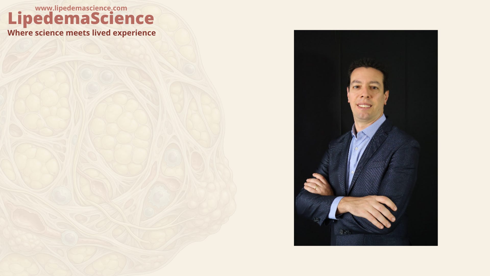
Dr. Anderson Nadiak Bueno
Vascular Surgeon and Vascular Ultrasound Specialist
CRM-SP: 122.105 | RQE: 46.614 and 46.614-1
Researcher
Vascular Ultrasound Residency Supervisor, FMABC
Scientific Director, LIPCOR
São Paulo, Brazil
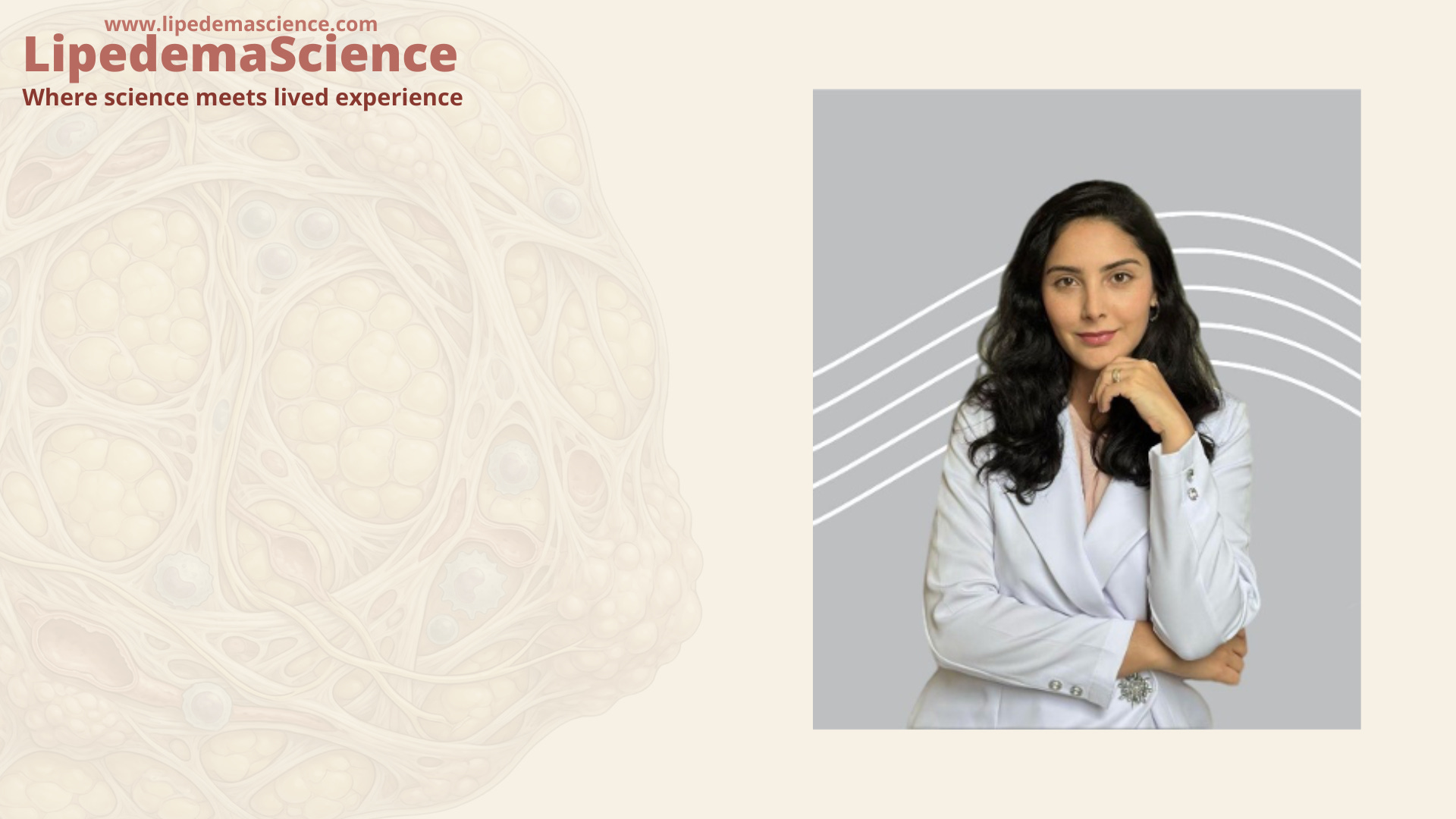
Dr. Thais Oliveira Foureaux
Radiologist
CRM-SP: 222.354 | RQE: 100.849
Researcher
Director of Scientific Production, LIPCOR
São Paulo, Brazil
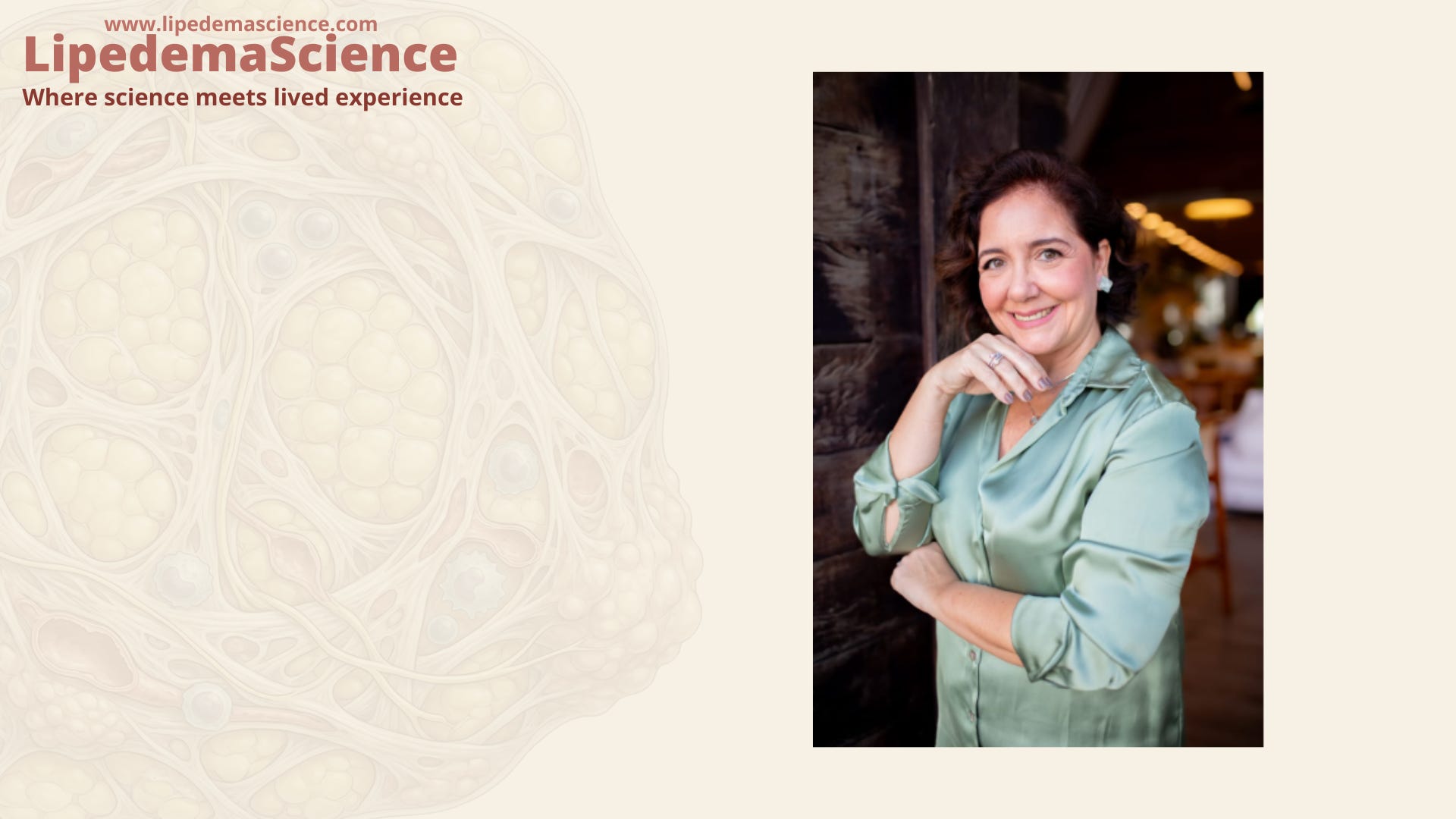
Dr. Dayana Ornelas
Radiologist
CRM-BA: 15.043 | RQE: 12.880 / 17.975
Researcher
Director of Scientific Integration, LIPCOR
Salvador, Brazil
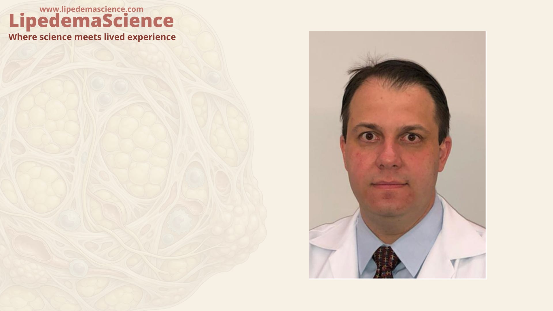
Dr. Alexandre Sacchetti Bezerra
Vascular and Endovascular Surgeon
CRM-SP: 112.815 | RQE: 28.114 / 28.115
Researcher
Professor of Vascular Surgery, FMABC
Full Member of the Brazilian Society of Angiology and Vascular Surgery (SBACV)
São Paulo, Brazil
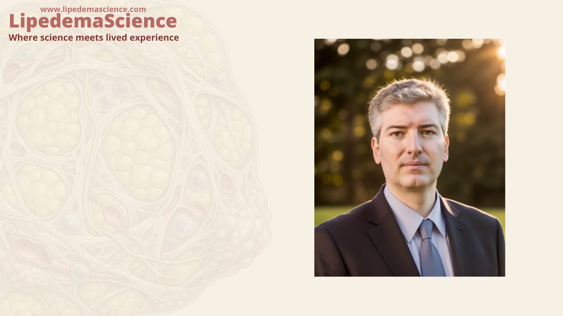
Dr. Alexandre Campos Moraes Amato, MD, PhD
Vascular and Endovascular Surgeon
CRM-SP: 108.651 | RQE: 29.069
Researcher
Amato Duo
São Paulo, Brazil