Turmeric, ginger, berries, extra virgin olive oil and oily fish are often described as anti-inflammatory foods. Many of them contain nutrients or bioactive compounds that are genuinely interesting from an inflammation perspective. Adding one of them to a meal, however, does not tell us whether the diet as a whole is anti-inflammatory, and it tells us even less about the wider conditions in which the body is living.
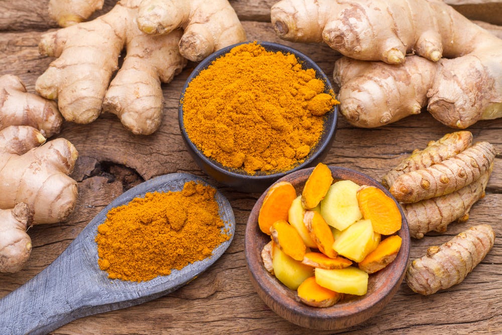
This distinction matters in lipedema because nutrition advice can quickly become a search for individual foods to add or remove. Turmeric is added because of curcumin, berries because of polyphenols, oily fish because of EPA and DHA. At the same time, sleep, stress, pain, physical activity, other illnesses and the broader dietary pattern may receive much less attention.
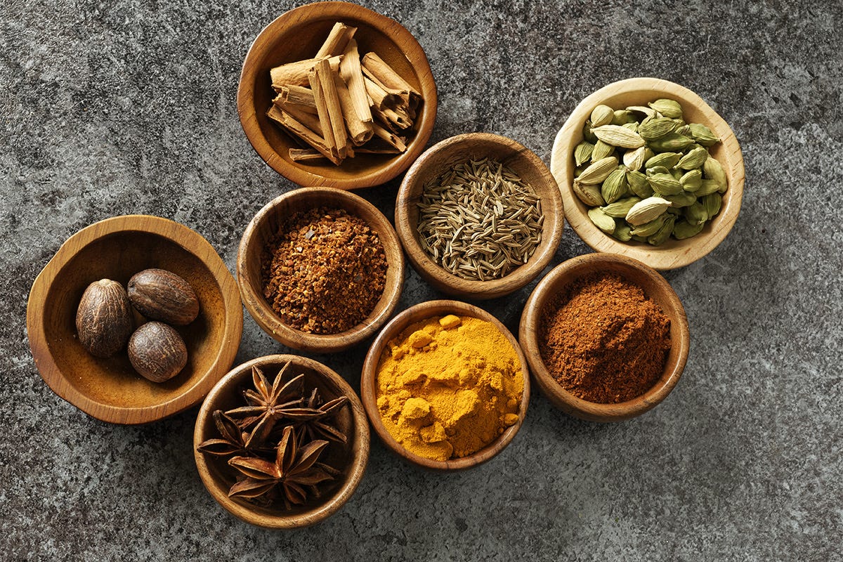
I find it more useful to separate the subject into three levels. The first is the individual food or compound. The second is the dietary pattern repeated over time. The third is the wider lifestyle and living conditions surrounding both. The levels overlap, but the evidence behind them is not interchangeable.

Anti-inflammatory foods are only the first level
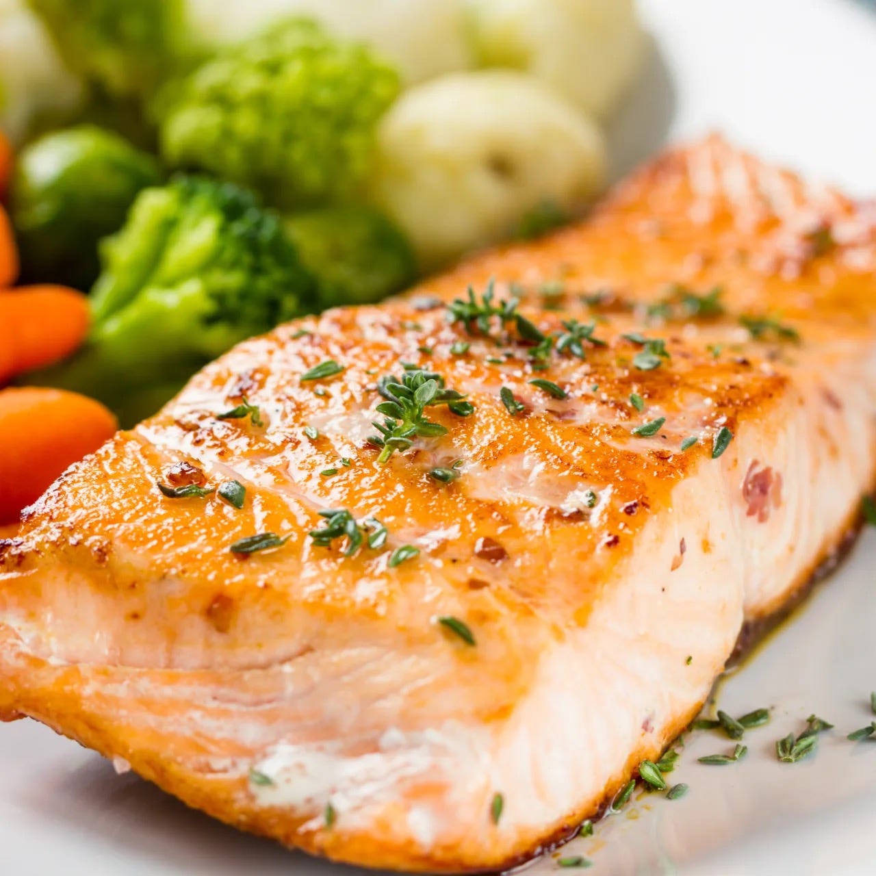
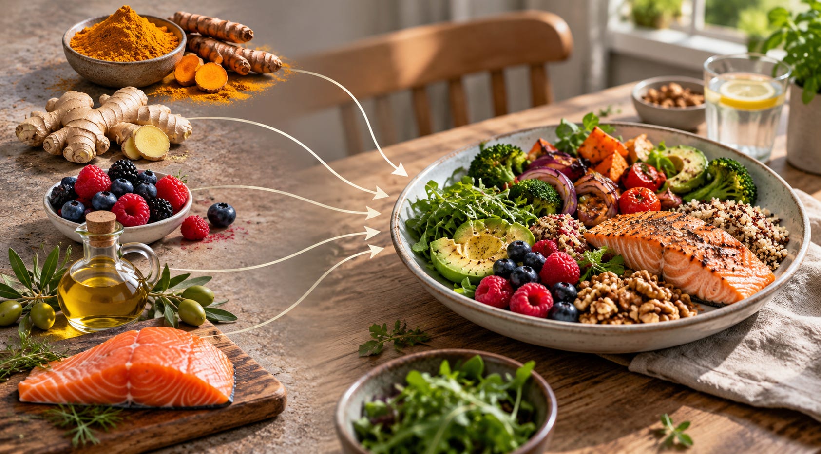
Individual foods can contain compounds that influence inflammatory biology. Oily fish provides the long-chain omega-3 fatty acids EPA and DHA. Extra virgin olive oil contains phenolic compounds. Berries contain several classes of polyphenols, while turmeric provides curcuminoids.
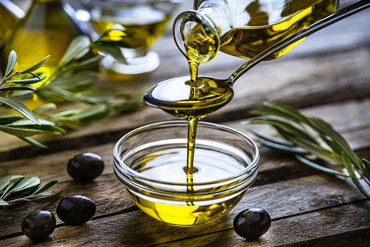
The strength of the human evidence differs considerably between them. EPA and DHA have been studied extensively in humans. For many plant and spice compounds, a larger part of the literature comes from laboratory experiments, small clinical studies or concentrated preparations that bear little resemblance to the amount normally eaten in food.
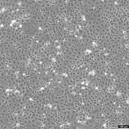
That difference matters to me because of my own laboratory background. My master’s research in food science covered inflammation, bioactive peptides and gut health, using simulated gastrointestinal digestion, human intestinal Caco-2 cell models and IL-8 ELISA measurements. Working with these models makes the distance between a biological mechanism and a human health outcome very tangible.
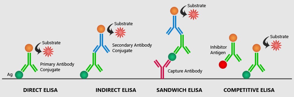
A compound can alter inflammatory signalling in cultured cells without demonstrating that a normal serving of the food containing it reduces pain, improves physical function or changes the course of a disease. Mechanistic evidence can help explain how something might work. It does not automatically establish that it works in everyday life.
This is why I see individual anti-inflammatory foods as inputs into a larger dietary pattern rather than as treatments in themselves.
What the evidence says about an anti-inflammatory diet
The evidence becomes different when researchers study entire dietary patterns.
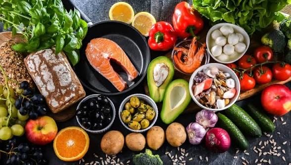
Mediterranean-style diets are one of the better studied examples. A systematic review and meta-analysis published online in 2025 and in the September 2026 issue of Nutrition Reviews included 33 randomized controlled trials with 3,476 participants. Mediterranean-style interventions reduced hs-CRP, IL-6 and IL-17 compared with control diets, while conventional CRP, IL-10, TNF-alpha and total antioxidant capacity did not change significantly. Subgroup results also varied according to characteristics such as age, intervention duration and cardiovascular disease status.
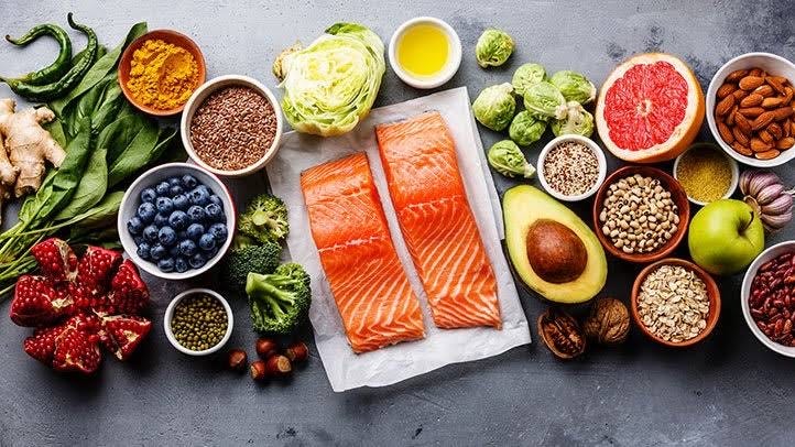
Those findings are more informative than simply calling the Mediterranean diet “anti-inflammatory.” Some inflammatory markers changed while others did not, and the effects were not identical across participants.
A smaller randomized trial provides another example. In the AUSMED Heart Trial, people with coronary heart disease were assigned to a Mediterranean or low-fat dietary intervention for six months. Among the 56 participants who completed the trial, the Mediterranean diet produced a significantly lower Dietary Inflammatory Index score than the low-fat diet, but the differences in hs-IL-6 and hs-CRP were not statistically significant.
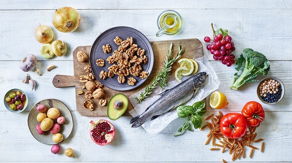
A related analysis of the same trial found that greater improvement in the Dietary Inflammatory Index correlated with a larger reduction in hs-IL-6, although the sample was small.
Taken together, these studies support the idea that dietary patterns can influence some inflammatory measures. They also show why the word anti-inflammatory should not be treated as an on-off switch.
What you replace matters too
Dietary research has another complication that is often missing from social-media discussions. Changing a diet rarely means adding one food while everything else remains untouched.
If someone begins eating more whole grains, refined grains may decrease. If fish appears more often, it replaces another main course. Increasing legumes, vegetables or nuts changes the proportion of other foods in the diet.
This means that part of an observed effect may come from what is removed or displaced rather than from the food being added in isolation.
A 2020 systematic review and meta-analysis of randomized trials compared whole grains with refined grains or placebo. The pooled result suggested a small reduction in CRP, with the confidence interval reaching the boundary of no effect. The authors found some additional cardiometabolic differences but did not consider the evidence strong enough to support broad treatment claims for cardiovascular disease. The comparison is informative precisely because it is a replacement comparison, not a study of simply adding one “healthy” food.
That replacement effect is one reason I am cautious when a particular ingredient is credited with the effect of an entire dietary pattern.
What about inflammatory foods?
The same caution is needed when foods are placed on the opposite list and described as inflammatory.
Ultra-processed food is a useful example. A 2025 cross-sectional analysis of 9,254 adults in NHANES examined ultra-processed food intake as a proportion of total energy intake. Compared with the lowest intake category, some higher categories had a greater adjusted likelihood of elevated hs-CRP, but the association was not a simple stepwise dose-response. Because the study was cross-sectional, it cannot establish that ultra-processed food caused the higher inflammatory marker. Other characteristics associated with the overall dietary pattern may still contribute.
The distinction is important. A habitual diet in which a large proportion of energy comes from ultra-processed food is one research question. Whether eating one processed food on Saturday afternoon causes a meaningful inflammatory state is another.
The same logic works in both directions. A serving of blueberries does not make an otherwise poor dietary pattern anti-inflammatory, and one ice cream does not define an otherwise nutritious diet.
Sleep shows why the wider context matters
Food is consumed by a body that has also slept, moved, recovered and experienced stress. Those factors do not sit in separate biological compartments.
Sleep research provides a particularly useful example because pain and inflammation do not always move together.

In a 2025 randomized crossover study, 39 healthy adults shortened their sleep substantially for two nights. Participants rated some experimental heat and cold pain as more painful after sleep restriction, while the inflammatory plasma markers measured in the study did not significantly change. The authors also noted poor adherence to the intended sleep-restriction condition, which is an important limitation.
A broader meta-analysis adds a different time scale. It included 35 experimental studies and 887 participants and found that multiple nights of partial sleep deprivation, around 4.3 hours of sleep for at least three nights, were associated with increases in circulating IL-6 and CRP. A single night of total or partial sleep deprivation was not associated with the same inflammatory changes. The paper was published online in 2025 and appears in the 2026 volume of the Journal of Sleep Research.
These findings help illustrate why “I hurt more today” and “my systemic inflammation increased today” are not equivalent statements. Duration, outcome and context matter.
Stress is relevant, but the evidence has limits
Psychological stress can also affect inflammatory signalling, although it is easy to overextend this literature.

A meta-analysis of 34 laboratory studies found increases in circulating IL-1 beta, IL-6, IL-10 and TNF-alpha following acute psychological stress. CRP did not significantly increase in that analysis.
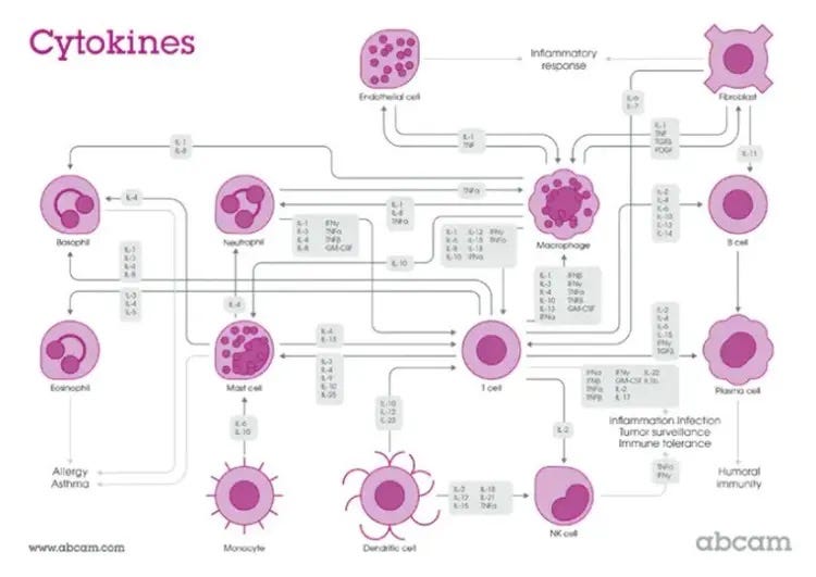
These were acute experimental stressors. They cannot simply be translated into claims about months of grief, job pressure or caregiving, and they do not establish a lipedema-specific mechanism.
They do, however, reinforce a broader point. Food is only one exposure occurring within a much larger physiological context.
What my own history has made me question
My interest in this subject is also personal.
My lipedema symptoms began around the age of 15, and the physical changes were documented by my general practitioner. My late teens and early twenties also included a serious illness at 18 and a major family bereavement at 20. Around that broader period, my pain became severe enough that I repeatedly sought help from my GP.
In 2018, while completing my master’s degree in food science, I experienced another particularly difficult period. My stress level was high, and my legs became substantially more swollen and painful.
Between 2021 and 2022, I had four lipedema surgeries involving my legs and arms. The procedures improved important aspects of my condition, particularly pain and mobility, but they did not remove the fact that I continue to live with lipedema through both easier and more demanding periods of life.
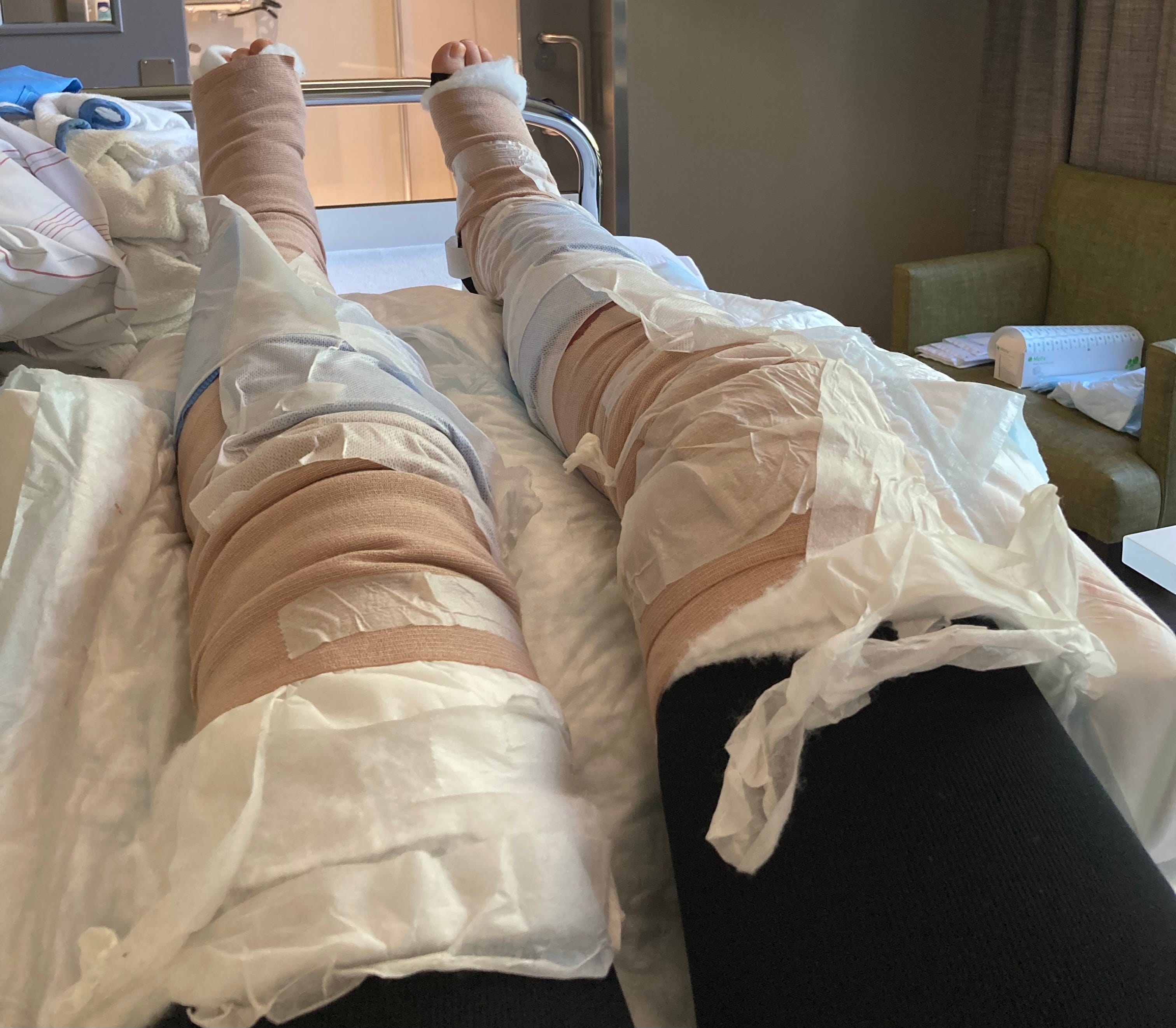
Looking backwards, I cannot separate all the variables involved in those changes. Hormones, illness, grief, sleep, activity, diet, pain and stress overlapped. My history cannot demonstrate which factor caused which symptom.
What it did allow me to do was notice patterns.
The factors I repeatedly noticed most strongly in my own body were hormonal changes, high sugar intake and stress. I treat those as observations from one person rather than evidence that the same triggers apply to everyone with lipedema.
My tolerance hypothesis
One observation has become particularly interesting to me.
When I sleep well, move regularly, eat well, recover and experience relatively little stress, I often seem to tolerate more. I can have chocolate, cake or ice cream without noticing an obvious flare in my symptoms.
During periods of greater stress, grief, illness or poor recovery, the same foods can appear to bother me more.
Before turning that into a biological theory, several simpler explanations have to be considered. Poor sleep can increase pain sensitivity. Stress can change appetite, eating behaviour and pain perception. Menstrual phase, portion size, earlier meals, physical activity and expectations may differ between occasions.
After those explanations are considered, I think one further question is reasonable. Could the total physiological load a person is carrying alter the threshold at which another exposure becomes noticeable?
At present, I regard that as a personal hypothesis. It has not been demonstrated as a mechanism in lipedema.
That distinction allows lived experience to generate questions without allowing it to become proof.
Is lipedema itself an inflammatory disease?
This is where the discussion becomes more complicated.
Inflammation is frequently mentioned in lipedema, sometimes as though the condition has already been established as a uniform systemic inflammatory disease. The available research does not support such a simple conclusion.
A 2023 tissue study examined subcutaneous adipose tissue from 32 women with lipedema and 14 age- and BMI-matched controls. The researchers found larger adipocytes in affected thigh tissue and a trend toward increasing fibrosis with stage. Macrophage markers were increased in affected thigh tissue, and the macrophage profile showed a transient shift toward an M2-like phenotype. Later stages showed features consistent with a more pro-inflammatory profile.
This is a local tissue finding. It does not automatically tell us what is happening systemically.
Blood studies provide another perspective. A 2026 retrospective study compared 78 women with lipedema, 76 women with obesity without lipedema and 75 normal-weight controls. CRP and erythrocyte sedimentation rate were higher in both the lipedema and obesity groups than in normal-weight controls, while there was no significant difference between the lipedema and obesity groups for those measures. BMI was substantially higher in both groups than in the normal-weight controls, which makes adiposity an important confounder.
A much smaller study comparing 13 women with lipedema with 13 age- and BMI-matched controls also found differences in circulating inflammatory and oxidative-stress-related proteins, alongside metabolic differences.
The tissue findings and blood findings therefore answer different questions. They also show why it is premature to describe every person with lipedema as being in one uniform systemic inflammatory state.
Nothing in these studies demonstrates that eating individual anti-inflammatory foods treats lipedema.
Testing my own assumption about lipedema and obesity
I also think it is important to test the assumptions we bring into a subject.
I started with the belief that lipedema is biologically distinct from ordinary obesity. There are good clinical reasons to distinguish them, and reducing lipedema to excess weight has contributed to misunderstanding and weight stigma.
At the same time, that starting position cannot be protected from evidence.
Lipedema and obesity can coexist. Some local tissue findings appear different from patterns commonly described in obesity, while some systemic inflammatory measurements overlap considerably. BMI, adiposity, disease stage and tissue location can all influence what researchers measure.
The more useful research question is therefore which biological features are genuinely specific to lipedema, which overlap with obesity and which are influenced by other characteristics of the people being studied.
Current evidence does not settle that question.
Why the pyramid has three levels
The pyramid accompanying this article is intentionally not a ranking of which factor has the largest biological effect.
At the top are single foods, where evidence varies by compound, dose and outcome.
The middle represents the dietary pattern, including the foods eaten repeatedly and what those foods replace.
The broad base represents lifestyle and living conditions. This includes sleep, stress, movement, pain, other illnesses, finances and access to care.
The base is wider because it covers more of the environment in which a person lives, not because I am claiming that lifestyle has a mathematically larger effect than diet. Studies of sleep, diet, stress and other exposures use different populations and outcomes and cannot simply be placed on one common effect-size scale.

Time runs underneath the entire pyramid because these exposures are repeated over months and years.
I also include the tolerance idea separately and label it as my observation rather than an established mechanism.
Sustainability matters more than collecting anti-inflammatory ingredients
This broader model also changes what an anti-inflammatory lifestyle means.
Lifestyle is not only a collection of personal decisions. Pain can limit movement. Another illness can disrupt sleep and recovery. Financial circumstances shape access to food, treatment and exercise. Work and caregiving can reduce the time available for rest. Access to healthcare differs greatly between people and countries.
For someone with lipedema, a sustainable approach therefore has to fit inside a real life rather than an idealized health routine.
Needs can also change. A period of grief, illness or prolonged sleep loss may require more recovery and fewer additional demands than a stable period. The same person may have different capacity at different points in life.
This does not make one piece of cake a failure, and it does not make a spoonful of turmeric a treatment.
Individual foods can contribute useful nutrients and bioactive compounds. Dietary patterns provide a broader nutritional exposure over time. Sleep, stress, movement, pain, other illnesses and living conditions add further context. In lipedema, the role of inflammation itself remains an evolving research question, which makes it especially important to distinguish mechanistic plausibility, personal experience and evidence from human studies.
That wider view is the reason I no longer think “Which anti-inflammatory food should I add?” is the most useful question on its own.

About the author
CarinaW is the founder of LipedemaScience. She has a bachelor’s degree in Human Nutrition and a master’s degree in Food Science from the Norwegian University of Life Sciences. Her laboratory background includes research on inflammation, bioactive peptides and gut health, using simulated gastrointestinal digestion, human intestinal Caco-2 cell models and IL-8 ELISA measurements. She has also worked with food microbiology and whole-genome sequencing, including as a laboratory manager, and is a co-author of peer-reviewed research. Diagnosed with lipedema in 2012, she combines scientific training with long-term lived experience while keeping personal observations separate from research evidence.